Review the orientation of the subtalar joint axis. Because the subtalar joint's axis is oriented obliquely to the three reference planes, the joint's motion is triplanar. In an open chain, the calcaneus and forefoot move simultaneously.
In a closed chain, when body weight stabilizes the foot against the floor, subtalar joint motion requires motion in proximal segments and joints like the talus, tibia, and knee.
Let the ankle and foot hang over end of table. Place the calcaneus in the frontal plane by positioning the opposite hip in flexion, abduction and external rotation.
Measuring subtalar range of motion (Elveru,Rothstein, Lamb, & Riddle, 1988)
Use a skin pencil to draw a line that bisects the middle third of the calcaneus.Draw a second line that bisects the distal third of the leg. Center the goniometer's axis between the malleoli in the frontal plane. Align the goniometer's stationary arm so that it parallels the line on the leg's distal third. Align the moving arm so that it parallels the line on the calcaneus. "Lock the forefoot" by grasping the fourth and fifth metatarsal heads and moving them dorsally (Elveru,Rothstein, Lamb, & Riddle, 1988, p. 680 and Figure 4). Maximally supinate the subtalar joint, realign the goniometer's arms, and read the degrees of inversion. Maximally pronate the STJ, realign the goniometer, and read the degrees of eversion. | 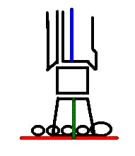
|
|---|
Finding subtalar joint neutral (STJN) by using two techniques
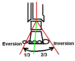
To find by a traditional (Root, Orien, Weed, & Hughes, 1971) definition:
|
|---|
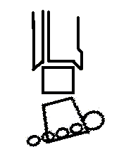
To find by palpation (Elveru,Rothstein, Lamb, & Riddle, 1988)
- Palpate medial and lateral aspects of the head of talus.
- Invert and evert the rearfoot in frontal the plane.
- The subtalar joint is neutral where the talar head protrudes equally on medial and lateral sides.
|
|---|
Find STJN by palpation while "gently dorsiflexing the ankle to the point of resistance" (Donatelli, 1990, p. 138) through the fourth and fifth metatarsal heads.
Compare the planes formed by (1) metatarsal heads and (2) the calcaneus' inferior surface (a line perpendicular to the calcaneal bisector that you drew earlier.
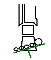
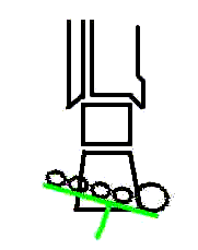
Forefoot varus Forefoot valgus
If the planes are not parallel, measure the difference with a goniometer and report it in degrees of forefoot varus or forefoot valgus. | |||||||||
|---|---|---|---|---|---|---|---|---|---|
- Find STJN by palpation.
- Passively dorsiflex the ankle while maintaining the STJ in neutral.
- Measure the ankle's PROM in dorsiflexion when the STJ is neutral and report in degrees.
When the ankle's passive dorsiflexion is limited, the cause can be structural (bony and fixed or rigid) or "functional," related to shortened soft tissue. Whatever the source of the limited ankle dorsiflexion, one might compensate by pronating the subtalar joint. Because this compensation can have untoward consequences, the therapist may seek to prevent it.
If the ankle's limited passive dorsiflexion relates to short gastrocnemeus and soleus, the person may increase isolated ankle dorsiflexion by stretching these muscles. Devise a technique that achieves this without simultaneously pronating the subtalar joint.
If the ankle's limited passive dorsiflexion relates to a structural or bony abnormality, approaches that focus on soft tissues are not relevant. Instead, the therapist may use a very simple orthosis: the heel wedge. Explain how a heel wedge, not more than 1/4" in height, can permit functional dorsiflexion while eliminating the need for compensatory pronation during midstance and terminal stance.