Each lab group needs:
- gait belt (one belt for each pair of students)
- summary of joint motion and muscle activity during the gait cycle
- Reviewing foundational information
Use the summary of joint motion and muscle activity to review the definitions of gait phases.
- Reviewing pelvic movements
Review the influence of pelvic rotation on lower extremity movement. You can demonstrate by rotating your pelvis in a transverse plane with both feet stable on the floor.
- Assessing pelvic movement while guarding the patient
Assess pelvic rotation and lateral pelvic drop while guarding at least three different partners:
- at the side, with one hand under a gait belt.
- from the rear, with hands on the gait belt at either side of pelvis.
Be alert for individual differences in pelvic rotation and lateral pelvic drop.
- Pelvic movement during (left) loading response
Discuss the pelvic movements that occur during loading response (roughly 0 to 10 percent of the gait cycle), when the pelvis drops on the contralateral side, and begins to rotate forward on the opposite side. Focus on left loading response and determine:
- which joints must move for this pelvic motion to occur?
- what motions occur at those joints?
- what forces, including muscular forces, produce these joint motions?
Discuss this problem for five or ten minutes, then compare your conclusions with the commentary at the end of the lab procedure.
- Facilitating appropriate gluteal activity while guarding the patient
Now that you've investigated the forces that contribute to normal pelvic movement, return to the guarding techniques you practiced earlier.
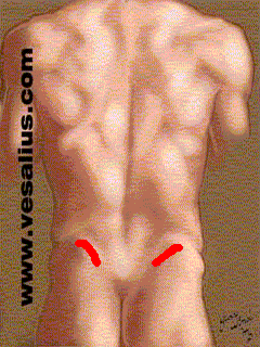
Unless your patient is so unstable that he or she requires extremely careful guarding, you can move your hands from the gait belt, and employ a technique that is appropriate for facilitating movement and muscle activity in many patients. Standing behind your partner, use your fourth and fifth fingers to apply downward pressure on the gluteus medius at those times during the gait cycle when the hip abductors are active.
You should initiate your pressure at initial contact, and sustain it through midstance. The pressure should be directed largely downward, with relatively small anterior and medial components. A good practice is to aim your pressure at that spot on the floor where the foot's center of pressure should be. You can perform this facilitation even as you maintain hold of the gait belt.

- Facilitating appropriate paraspinal activity while guarding the patient
You can combine the gluteus medius facilitation with another that is directed at the "paraspinal" muscles (the muscles that are "near the spine," that is, the erector spinae and transversospinalis groups). To understand the paraspinals' timing during the gait cycle, palpate them on yourself by placing the fingers of either hand on both sides of the midline of your back. Assess the paraspinals' activity as you walk. Correlate what you feel with information on muscle activity.
- Refining the pelvic girdle facilitation
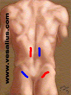
Working again with your partner, facilitate the gluteus medius as you did before, but combine that pressure with simultaneous pressure, by means of the thumb, on the contralateral paraspinal muscles.
Do this by spreading your hand as wide as possible so that:
- the fourth and fingers rest on the gluteals and
- the thumbs rest on the paraspinals
During left loading response, as an example, simultaneously facilitate the left gluteals and the right paraspinals . During right loading response, simultaneously facilitate the right gluteals and the left paraspinals.
You will be most successful if you keep your hands and upper extremities fairly stiff and generate the movement in your own shoulders and trunk.

- Further refinements of pelvic girdle and trunk facilitation
Using the same hand position, you can cue your partner to alter the lumbar posture in flexion or extension. Do this by touching the lateral trunk with your second and third fingers, and applying gentle traction in various directions. You can facilitate a lateral weight shift by using your thumb to provide a gentle cue to the paraspinal muscles on one side. Practice these facilitations as your patient stands still.
- Facilitating through the shoulder girdle
You can facilitate the walking pattern from the shoulder girdle instead of the pelvis. After watching a demonstration, practice the facilitation, using the following "coaching points."
- Position the thumb (1) at the base of the scapular spine, and the second and third fingertips on the clavicle's anterior surface. Do not contact the top of the shoulder.


- Abduct your shoulders so that your elbows stay up.
- By dropping your elbow (just a little), you will apply pressure to the scapular spine in a downward and slightly anterior direction, at the same time that you apply traction to the clavicles in the direction of backward rotation. If you can spread your hand sufficiently, try to contact the lateral aspect of the shoulder with your fourth or fifth fingertips. You can apply gentle traction on the skin in the direction of external rotation. The combination of these cues will facilitate a more erect or extended posture throughout the thoracic spine and shoulder girdle.
- To help the person start walking, cue a lateral weight shift by pressing medially on one scapula. This lateral shift places the weight on one leg, and frees the other leg so that it can swing forward.
- As you walk with the patient, concentrate on cueing extension of the stance side by using the combination of cues described above: press down, forward, and medially on the base of the scapular spine on the stance side as you apply traction in appropriate directions to the clavicle and lateral shoulder. Once again, this is easiest if you simply drop your elbow on the stance side.
- You can also facilitate the appropriate counter-rotation between the shoulder girdle and pelvis, but be very subtle; a little goes a long way.
Commentary - movement and muscle activity during left loading response
- Joints that must move to permit pelvic movement:
- hip joint
- lumbar intervertebral joints
- Joint motions that occur during left loading response:
Right pelvic drop involves:
- left sidebending of the lumbar intervertebral joints
- adduction of the left (loading response) hip
Right forward pelvic rotation involves:
- rightward rotation of the lumbar intervertebral joints
- internal rotation of the left (loading response) hip
In summary,
the left hip adducts and rotates internally,
while the lumbar intervertebral joints sidebend to the left and rotate to the right.
- Forces that produce these motions:
Gravity and inertial forces produce hip adduction during loading response. Consequently, hip abductors, particularly the gluteus medius and minimis, are active.
If we consider only the frontal plane, it is obvious that the the stance side abductors act eccentrically to control pelvic drop. However, if we consider that the hip simultaneously rotates internally in the transverse plane, the actions of the gluteus medius and minimis are less clear. The gluteus minimus and the anterior two-thirds of the gluteus medius are internal rotators. When we account for the muscles' actions in both the frontal and transverse planes, we conclude that they act:
concentrically, eccentrically, or isometrically (choose one).
The gravitational and inertial forces that produce right pelvic drop also cause the spine to sidebend to the left. During this lab session, you will palpate the paraspinal muscles and find that they are very active on the right during left loading response. Because the right erector spinae produce right sidebending and right rotation, it seems clear that the right erector spinae's action is eccentric when we consider just the frontal plane movement, that is, left sidebending. However, if we consider simultaneously the movements in both the frontal and sagittal plane, we might conclude that the erector spinae's action is nearly isometric, similar to the action we've hypothesized in the gluteus medius and minimus.
During this lab session, we learn a facilitation that seeks to encourage this simultaneous activity in the gluteals (on the loading response side) and the erector spinae (on the opposite, or preswing side).
Last updated 4-2-02 Dave Thompson PT
Use the summary of joint motion and muscle activity to review the definitions of gait phases.
Review the influence of pelvic rotation on lower extremity movement. You can demonstrate by rotating your pelvis in a transverse plane with both feet stable on the floor.
Assess pelvic rotation and lateral pelvic drop while guarding at least three different partners:
- at the side, with one hand under a gait belt.
- from the rear, with hands on the gait belt at either side of pelvis.
Be alert for individual differences in pelvic rotation and lateral pelvic drop.
Discuss the pelvic movements that occur during loading response (roughly 0 to 10 percent of the gait cycle), when the pelvis drops on the contralateral side, and begins to rotate forward on the opposite side. Focus on left loading response and determine:
- which joints must move for this pelvic motion to occur?
- what motions occur at those joints?
- what forces, including muscular forces, produce these joint motions?
Discuss this problem for five or ten minutes, then compare your conclusions with the commentary at the end of the lab procedure.
Now that you've investigated the forces that contribute to normal pelvic movement, return to the guarding techniques you practiced earlier.
Unless your patient is so unstable that he or she requires extremely careful guarding, you can move your hands from the gait belt, and employ a technique that is appropriate for facilitating movement and muscle activity in many patients. Standing behind your partner, use your fourth and fifth fingers to apply downward pressure on the gluteus medius at those times during the gait cycle when the hip abductors are active.You should initiate your pressure at initial contact, and sustain it through midstance. The pressure should be directed largely downward, with relatively small anterior and medial components. A good practice is to aim your pressure at that spot on the floor where the foot's center of pressure should be. You can perform this facilitation even as you maintain hold of the gait belt. | 
|
|---|
You can combine the gluteus medius facilitation with another that is directed at the "paraspinal" muscles (the muscles that are "near the spine," that is, the erector spinae and transversospinalis groups). To understand the paraspinals' timing during the gait cycle, palpate them on yourself by placing the fingers of either hand on both sides of the midline of your back. Assess the paraspinals' activity as you walk. Correlate what you feel with information on muscle activity.
Working again with your partner, facilitate the gluteus medius as you did before, but combine that pressure with simultaneous pressure, by means of the thumb, on the contralateral paraspinal muscles.
Do this by spreading your hand as wide as possible so that:
During left loading response, as an example, simultaneously facilitate the left gluteals and the right paraspinals . During right loading response, simultaneously facilitate the right gluteals and the left paraspinals. You will be most successful if you keep your hands and upper extremities fairly stiff and generate the movement in your own shoulders and trunk. | 
|
|---|
Using the same hand position, you can cue your partner to alter the lumbar posture in flexion or extension. Do this by touching the lateral trunk with your second and third fingers, and applying gentle traction in various directions. You can facilitate a lateral weight shift by using your thumb to provide a gentle cue to the paraspinal muscles on one side. Practice these facilitations as your patient stands still.
You can facilitate the walking pattern from the shoulder girdle instead of the pelvis. After watching a demonstration, practice the facilitation, using the following "coaching points."
- Position the thumb (1) at the base of the scapular spine, and the second and third fingertips on the clavicle's anterior surface. Do not contact the top of the shoulder.


- Abduct your shoulders so that your elbows stay up.
- By dropping your elbow (just a little), you will apply pressure to the scapular spine in a downward and slightly anterior direction, at the same time that you apply traction to the clavicles in the direction of backward rotation. If you can spread your hand sufficiently, try to contact the lateral aspect of the shoulder with your fourth or fifth fingertips. You can apply gentle traction on the skin in the direction of external rotation. The combination of these cues will facilitate a more erect or extended posture throughout the thoracic spine and shoulder girdle.
- To help the person start walking, cue a lateral weight shift by pressing medially on one scapula. This lateral shift places the weight on one leg, and frees the other leg so that it can swing forward.
- As you walk with the patient, concentrate on cueing extension of the stance side by using the combination of cues described above: press down, forward, and medially on the base of the scapular spine on the stance side as you apply traction in appropriate directions to the clavicle and lateral shoulder. Once again, this is easiest if you simply drop your elbow on the stance side.
- You can also facilitate the appropriate counter-rotation between the shoulder girdle and pelvis, but be very subtle; a little goes a long way.
- hip joint
- lumbar intervertebral joints
- Right pelvic drop involves:
- left sidebending of the lumbar intervertebral joints
- adduction of the left (loading response) hip
- rightward rotation of the lumbar intervertebral joints
- internal rotation of the left (loading response) hip
Right forward pelvic rotation involves:
In summary,
- the left hip adducts and rotates internally,
while the lumbar intervertebral joints sidebend to the left and rotate to the right.
If we consider only the frontal plane, it is obvious that the the stance side abductors act eccentrically to control pelvic drop. However, if we consider that the hip simultaneously rotates internally in the transverse plane, the actions of the gluteus medius and minimis are less clear. The gluteus minimus and the anterior two-thirds of the gluteus medius are internal rotators. When we account for the muscles' actions in both the frontal and transverse planes, we conclude that they act:
The gravitational and inertial forces that produce right pelvic drop also cause the spine to sidebend to the left. During this lab session, you will palpate the paraspinal muscles and find that they are very active on the right during left loading response. Because the right erector spinae produce right sidebending and right rotation, it seems clear that the right erector spinae's action is eccentric when we consider just the frontal plane movement, that is, left sidebending. However, if we consider simultaneously the movements in both the frontal and sagittal plane, we might conclude that the erector spinae's action is nearly isometric, similar to the action we've hypothesized in the gluteus medius and minimus.
During this lab session, we learn a facilitation that seeks to encourage this simultaneous activity in the gluteals (on the loading response side) and the erector spinae (on the opposite, or preswing side).