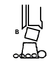
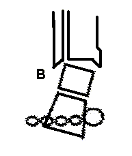
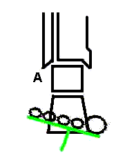
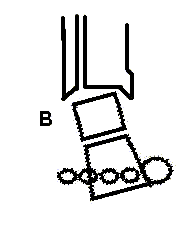
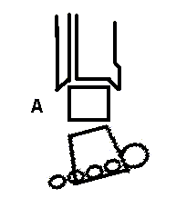
In uncompensated rearfoot varus (A), the calcaneus (and forefoot) are inverted when the subtalar joint is neutral.The person compensates (B) by pronating STJ to allow medial heel to touch the ground. | ||
---|---|---|
Compensation retards supination during midstance/terminal stance.The therapist can eliminate the need for compensation by supporting the deformity using an orthosis with a medial heel wedge. |
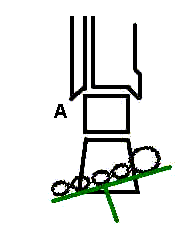
In uncompensated forefoot varus (A), the plane of metatarsal heads is inverted in relation to the rearfoot's plane when the subtalar joint is neutral.The person compensates (B) by pronating the STJ durng midstance and terminal stance to allow the first metatarsal to contact the ground. | |||||
---|---|---|---|---|---|
The compensation retards midstance/terminal stance supination.
The therapist can eliminate the need for compensation by supporting the deformity with an orthosis that incorporates a medial forefoot wedge. |
In uncompensated forefoot valgus (A), the plane of metatarsal heads is everted in relation to the rearfoot when the STJ is in neutral.The person compensates (B) by supinating the STJ during midstance then pronating during terminal stance. | |||||
---|---|---|---|---|---|
The compensation makes the foot less stable during preswing and push-off and, over time, produces joint laxity and pain.
The therapist can eliminate the need for compensation by supporting the deformity with an orthosis that incorporates a lateral forefoot wedge. |
When the ankle's passive dorsiflexion is limited, the cause can be structural (bony and fixed or rigid) or "functional," related to shortened soft tissue.Whatever the source of the limited ankle dorsiflexion, one can compensate by pronating the STJ during midstance and terminal stance. Because pronation has a sagittal plane component, dorsiflexion, the person can advance the tibia during stance by moving at the subtalar joint as well as the ankle. The compensatory pronation provides as much as 10 degrees of functional dorsiflexion (Tiberio, Bohannon, & Zito, 1989) but at the cost of rendering the foot mobile during push-off. |
---|
Rodgers, M.M. (1995). Dynamic foot biomechanics. Journal of Orthopedic and Sports Physical Therapy, 21, 306-316.
Tiberio, D. (1988). Pathomechanics of structural foot deformities. Physical Therapy, 68, 1840-9.
Tiberio, D., Bohannon, R.W., & Zito, M.A. (1989). Effect of subtalar joint position on the measurement of maximum ankle dorsiflexion. Clinical Biomechanics, 4, 189-191.