NQ-051a Answer: (C) Fornix
NQ-051b
Answer:
(B) Unilateral obstruction of
middle cerebral artery
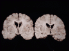
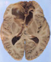
Pathology of the cases:
The area of necrosis is
indicated by the white arrows. The infarcted area includes the insula, the upper
temporal lobe and lower frontal-parietal lobe on one side. The pathologic
findings are most consistent with non-hemorrhagic infarction resulted from
unilateral obstruction of the middle cerebral artery (MCA).
[Website
on cerebral vascular territories]
|
|
Click on this icon to see the image.
|
Anterior cerebral artery: Infarction due to blockage of anterior cerebral artery (ACA) typically lead to infarction along the midline extending from the frontal tip and along the cingulate gyrus.
|
|
Click on this icon to see the image of a case of bilateral anterior
cerebral infarction. This infarct was resulted from blockage of
bilateral anterior cerebral artery. |
Venous infarction:
The brain has very good venous collaterals. Therefore, thrombosis must usually
be fairly extensive before an infarction can be resulted. Common sites are:
superior sagittal sinus (72%), lateral sinuses (70% combined), and straight
sinus (13%). Thrombosis commonly extends to several sinuses and veins.
Thrombosis of cerebral veins, especially the cortically ones, rarely results in
tissue damage, but thrombosis of the deep internal veins and the great vein of
Galen may cause severe damage to the basal ganglia and brain stem.
Macroscopically, the hemorrhagic infarct may extend from the thrombosed vein to
the white matter.
Venous
infarction usually leads to a triangular shaped infarction with the tip of the
triangle at the white matter.
-
The paravertebral venous plexus of Batson forms and extensive system of venous channels both within and alongside the spinal canal providing direct communication from peritoneal sites and the lower body to the cranial cavity. There are no valves in Batson’s plexus and flow may be bi-directional during the Valsalva maneuver or a change of body position. Venous infarction due to thrombosis of the Batson plexus is rather uncommon because of the large number of collaterals. The plexus of Batson is in the spinal cord so this answer is obviously wrong.
The anterior choroidal artery supplies an area medial to the
area under discussion.
[Online image of the anterior choroidal artery territory]