Case No.: A-007
Diagnosis: Herpes esophagitis
Organ: Esophagus
Last Updated: 1/21/2011
|
|
|
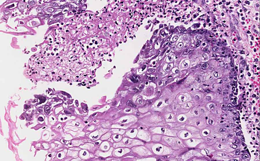
Hematoxylin & eosin |
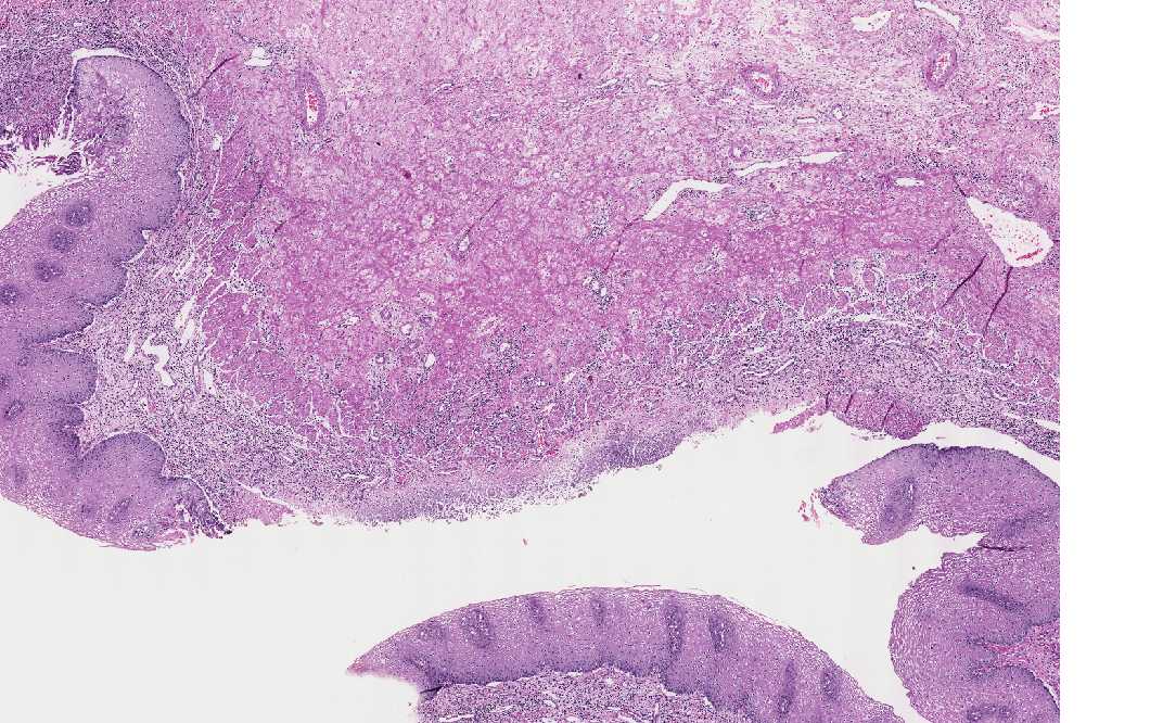
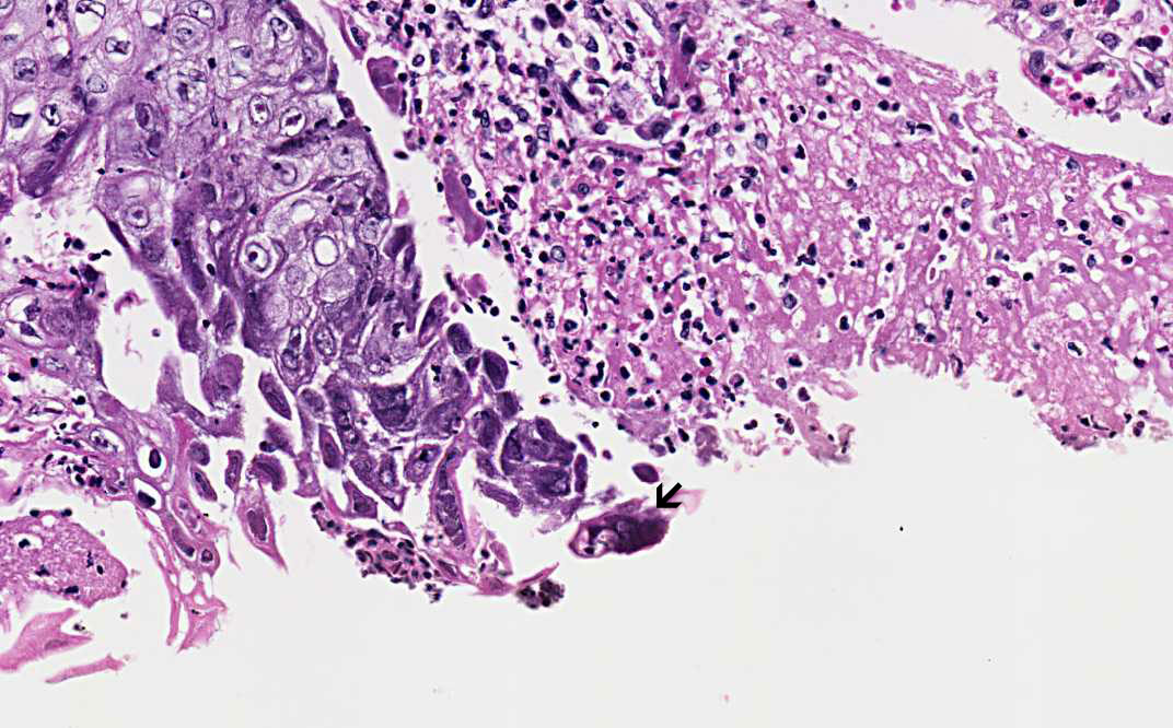
Area 1: Note that the ulcer is rimmed by intact mucosa. The ulcer is covered by a thin layer of fibrinous exudate with underlying granulation (contains a good number of neovascularization) associated with acute and chronic inflammation. Characteristically, the 3M cells (arrow) are noted in the squamous epithelium at the edge of the ulcer. It is, therefore, important to biopsy the edge of the ulcers. |
|
Hematoxylin & eosin |
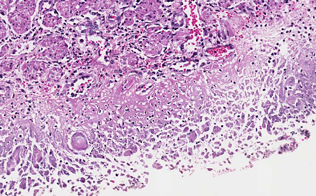
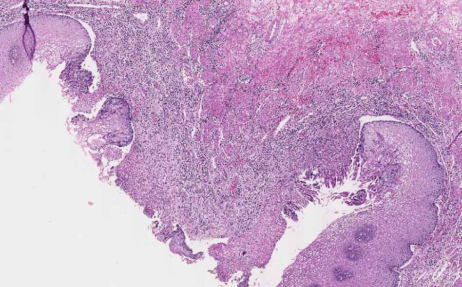
Area 2: The features are similar to that of Area 1 except that the 3M cells are more easily seen. |
|
History: The history of this case was not know as it was taken from the archival material. However, patients typically present with odynophagia and dysphagia. It is most often associated with immunocomprised conditions such as AIDS, recipients of solid organ transplantation, and post-chemotherapy treatment. It can, however, also occur occasionally in immune competent patients.
Clinical and Endoscopic Findings:
Histologic Highlights of this Case:
|
Bonus Images:
|
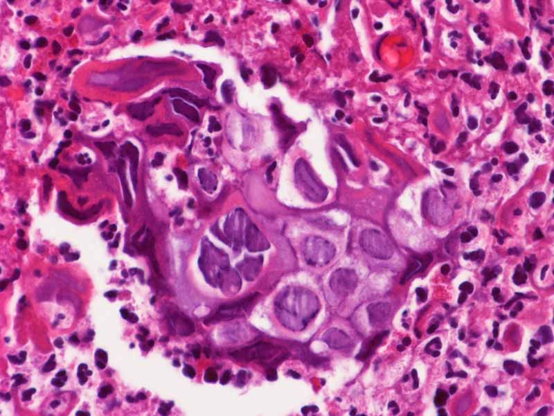
Hematoxylin & eosin |
High-magnification: This image is taken from a different case and at a higher magnification (60x). The 3M nuclei (molding, margination of chromatin, multinucleated) are well demonstrated. |
|
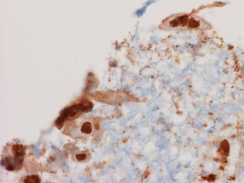
Immunohistochemistry |
Immunohistochemistry for HSV: Immunohistochemistry can help to detect some of the viral inclusion that would be difficult to be diagnosed by morphology. It provides good confirmation for the diagnosis. |
Original slide is contributed by James Fishback MD, Department of Pathology, Kansas University (Iowa Image Collection).