Case No.: A-015
Diagnosis: Well-differentiated neuroendocrine carcinoma (carcinoid) and atrophic gastritis with intestinal metaplasia
Organ: Stomach
Last Updated: 11/21/2011
|
|
|
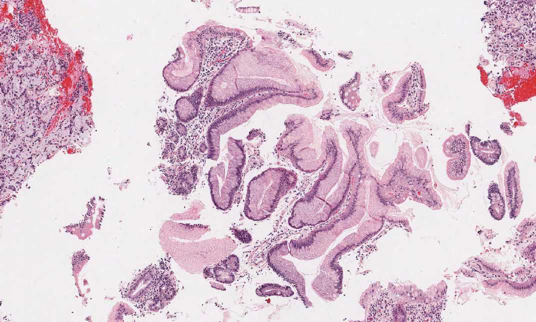
Hematoxylin & eosin |
Area 1: This is the area with gastric mucosa fragments. Note that intestinal metaplasia is present in some of the fragments. |
|
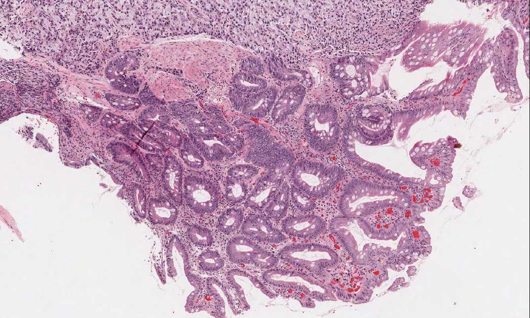
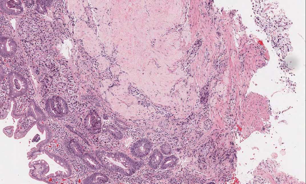
Hematoxylin & eosin |
Area 2: This is the area of residual gastric mucosa with chronic atrophic gastritis and intestinal metaplasia. |
|
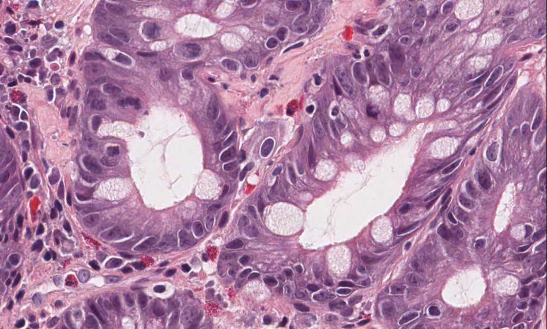
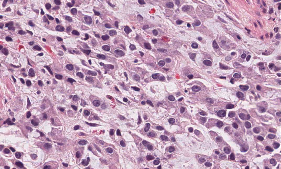
Hematoxylin & eosin |
Area 3: The overall feature of the tumor is that of a cellular neoplastic epithelial proliferation with a myxomatous stroma and no distinct glandular formation. The overall arrangement of the tumor cells and the nuclear features suggest an neuroendocrine carcinoma. |
|
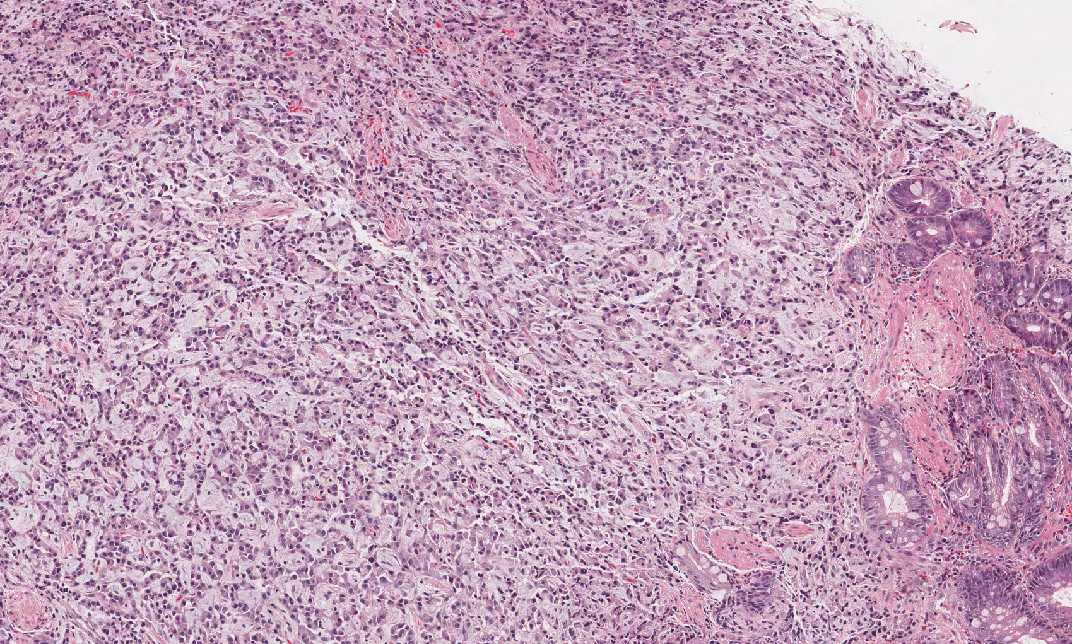
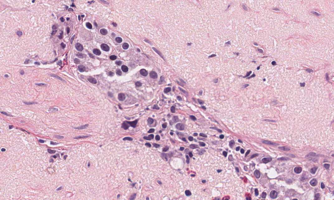
Hematoxylin & eosin |
Area 4: The tumor invades into the muscularis propria. |
|
History: This biopsy was taken from a 70 year-old woman. What is the organ? What is your diagnosis?
Histologic Highlights of this Case:
Comment:
|
Bonus Images:
|
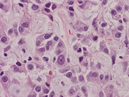
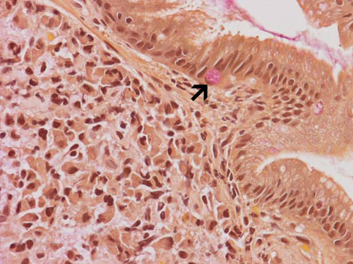
Hematoxylin & eosin |
High magnification: A high magnification image shows round, and rather monontonous nuclei with prominent nucleoli and "salt and pepper" like chromatin. Although some adenocarcinoma can have this kind of nuclei, the nuclear features raise a serious concern of neuroendocine tumor. |
|
Mucicarmine stain |
Mucicarmine stain: Although there is myxomatous stroma in this tumor, there is no genuine mucin production in the tumor cells. A mucicarmine stain demonstrates positive staining in the goblet cells (intestinal metaplasia) which serve as the internal control but not the tumor cell. |
|
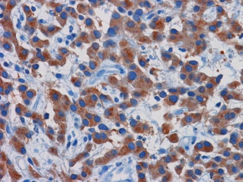
Synaptophysin |
Synaptophysin: Immunohistochemistry for synaptophysin confirms the neuroendocrine nature of this tumor. |
Original slide is contributed by Dr. Kar-Ming Fung, University of Oklahoma Health Science Center, Oklahoma, U.S.A.