|
History: The history of this case was not know as it was taken
from the archival material. The patient was an adult and has a history
of malignant hypertension. The specimens
were obtained at an autopsy.
Histologic Highlights of this Case:
-
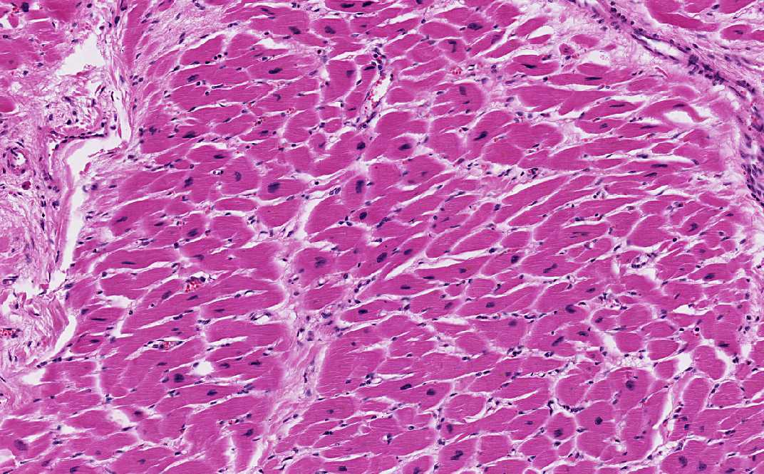
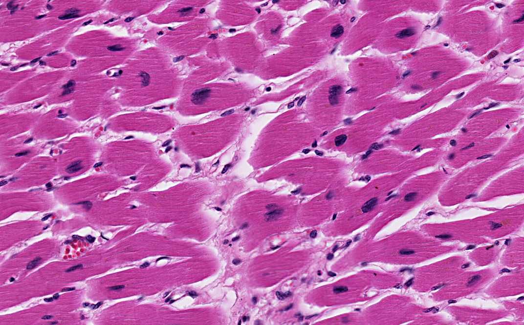
Heart: Patient with prolonged
hypertension, the heart is typically grossly hypertrophic featured
by concentric thickening of the left ventricular wall and increase
in weight. Microscopically, here is diffuse hypetrophy of the
cardiac muscle. The myocytes increases in width. The normal cardiac
myocyte is about 10-15 microns in with but in the hypertrophyic
myocytes, they can reach a width of 25 microns. The nuclei is also
enlarged and hyperchromatic. They often adopt a rectangular shape
and are termed "box-car nuclei". With the modern eyes, they do
resemble minivans or SUVs! (Heart- Area 1)
-
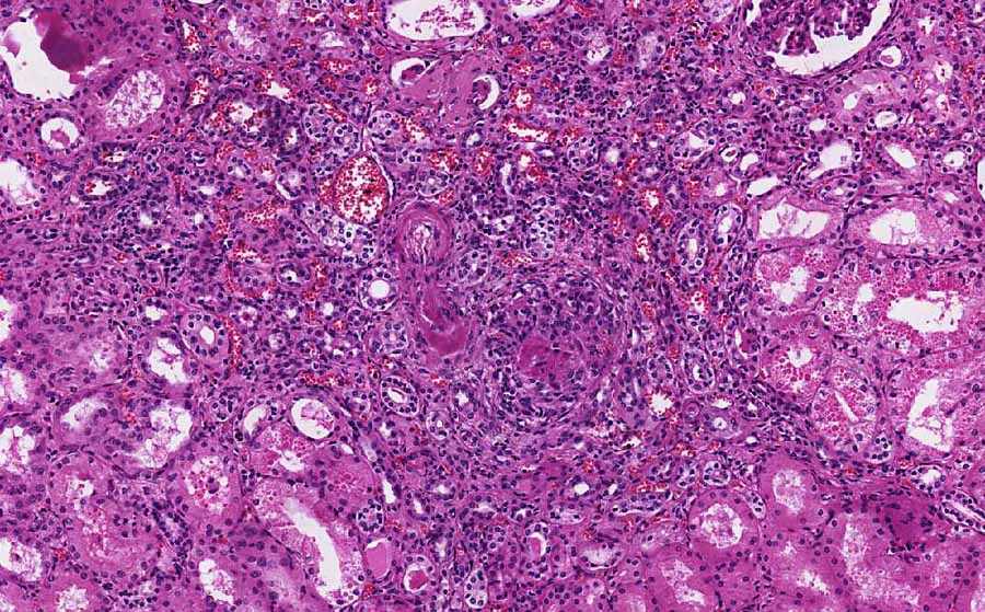
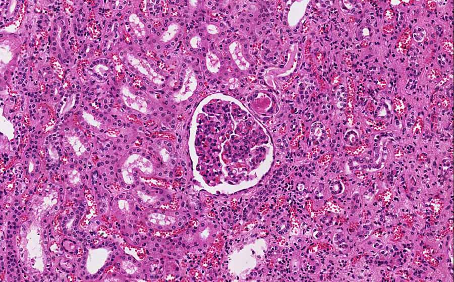
Kidney:
Malignant or
accelerated phase of hypertension will lead to accelerated
nephrosclerosis. Although this condition may develop in previously
normotensive individuals, it is often superimposed on preexisting
essential benign hypertension, other forms of non-malignant
hypertensions, and underlying chronic renal diseases particularly
glomerulonephritis and reflux nephtopathy. The longer the duration
of the disease the more likely to see a smaller kidney on gross
examination. The kidney may have a "flea-bitten"appearance featured
by small, pinpoint petechial hemorrhage on the cortical surface.
These petechia are resulted from rupture of arterioles or clomerular
capillaries of the kidney. The reason for this type of gross
pathologic changes may be explained by the presence of two
characteristic pathologic changes of blood vessels in malignant
hypertension:
·
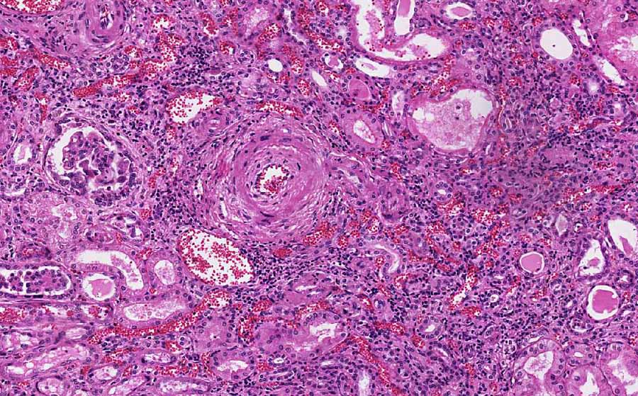
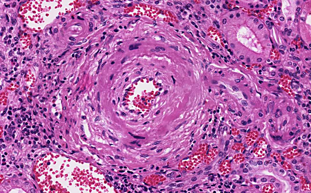
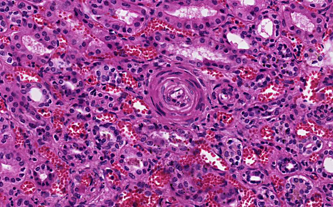
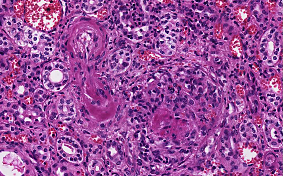
Concentric intimal thickening of blood vessels (hyperplastic
arteriolitis):
There is extensive intimal thickening due to a proliferation of
elongated, concentrically arranged smooth muscle cells and layers of
concentric collagen. Pale staining substance probably proteoglycans
and plasma proteins are also present in these concentric
thickenings. Histologically, these vessels resemble the cross
section of an onion (onin-skinning of vessels). (Kidney- Area 1 and
2)
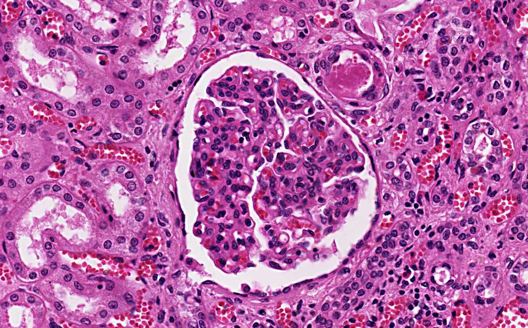
·
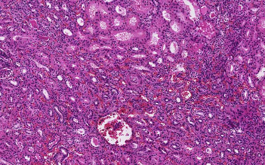
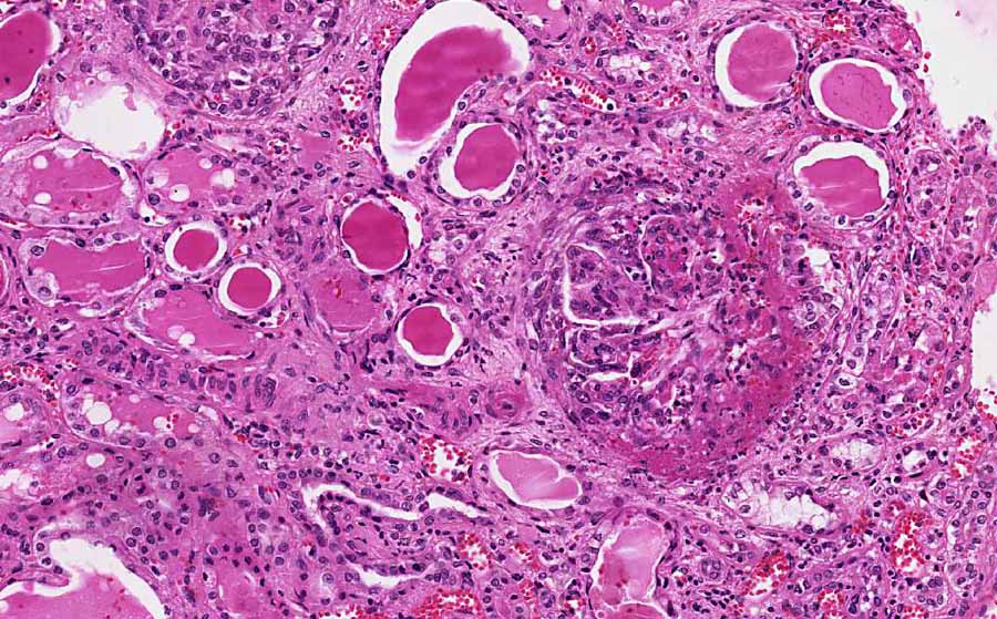
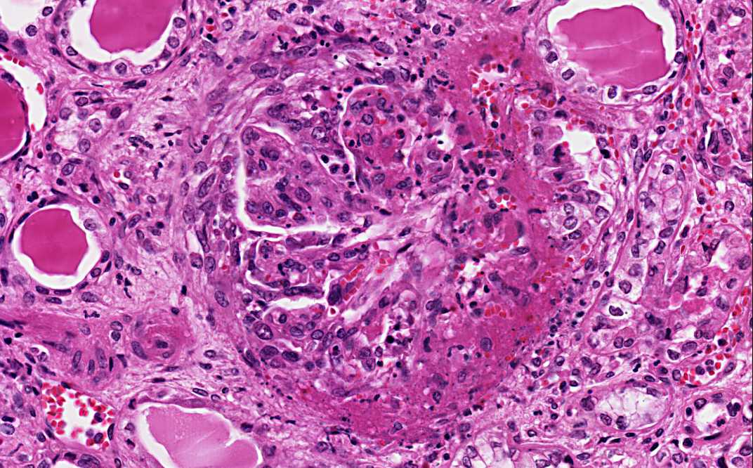
Fibrinoid necrosis of arterioles:
The blood vessel wall is partially or completely replaced by an
eosinophilic granular acellular fibrinoid substance (Kidney- Area 3)
which can be demonstrated by special stains or immunohistochemistry.
Sometimes the glomeruli may also become necrotic (Kidney- Area 4)
and infiltrated by neutrophils and the glomerular blood capillaries
may be thrombosed (Kidney- Area 5). Limited chronic
inflammatory cell infiltration may be associated with these vessels.
|