|
History: This slide was
retrieved from an archive and no history was available. This condition
often occurs in immune deficient patients, it can also occur in immune
competent patients as well as congenital form.
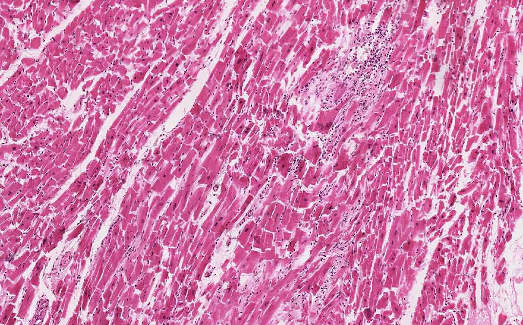
Histologic Highlights of this Case:
-
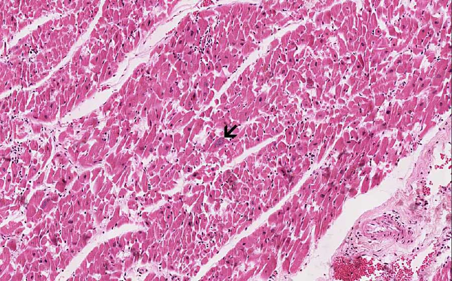
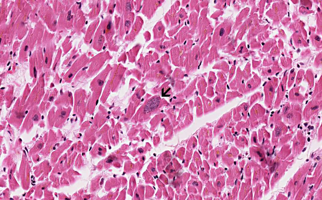
This specimen appears to be taken from a
subendocardial location as you can see the endocardium (arrow).
Based on its shape, it may well correspond to a papillary muscle.
-
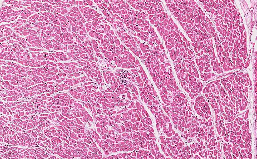
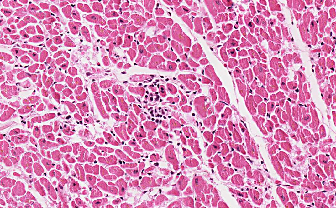
There is no area with necrosis,
fibrosis, or scar formation. The most obvious pathologic changes are
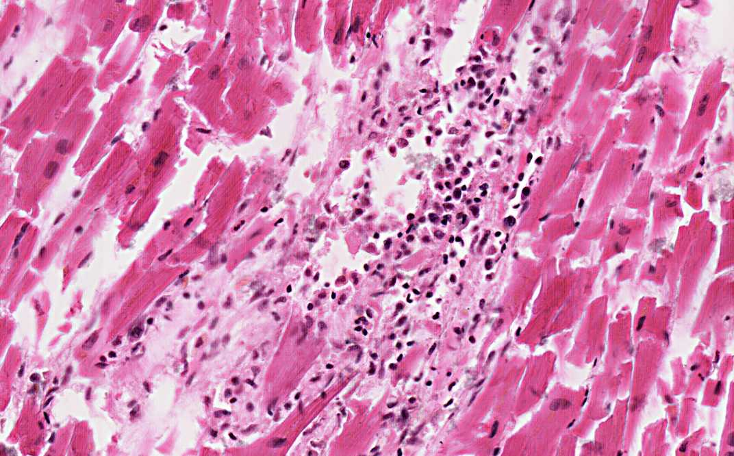
scattered small foci of inflammatory cell infiltration (Area 1 and
2). Some of the inflammatory foci are really small (Area 2).
Eosinophils are often but not always seen in Toxoplasma
myocarditis. No microorganisms appear to be associted with these
inflammatory foci.
-
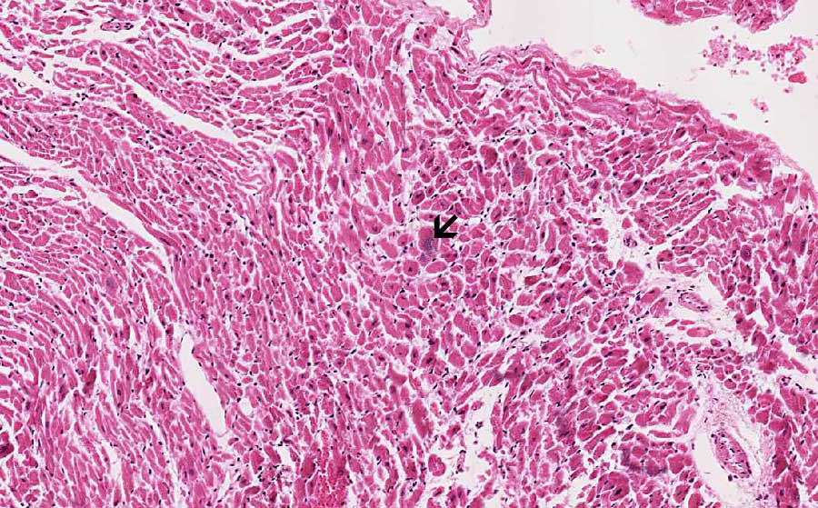
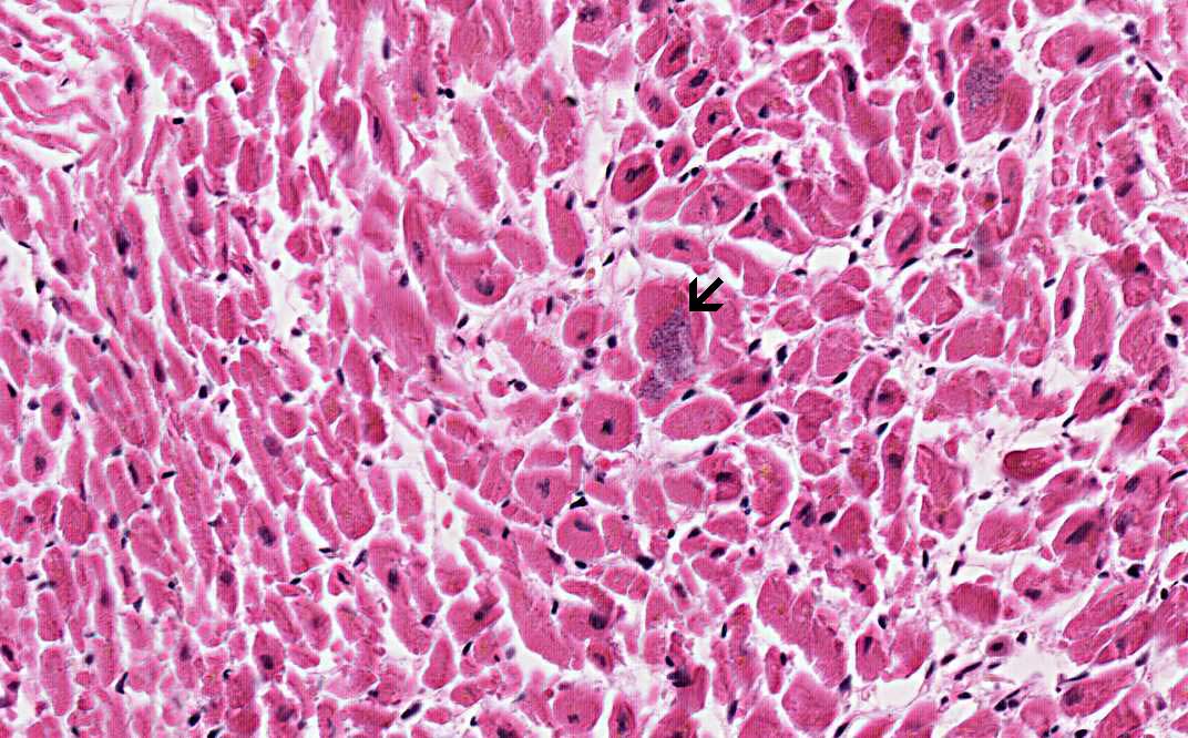
If you look at this slide more
carefully, however, you will see some large nucleus-like structures
inside the myocyte (Area 3 and 4). You would first think that these
are enlarged nuclei in hypertroophic myosites. However, they do not
look really like a nuclei and, instead, look like an
intracytoplasmic bag with many small dots. In addition, the myocytes
nearby do not have nuclei this big. This is rather unusual for
enlarged myocytes in myocardial hypertrophy. These structures are
instead the intracellular cysts (tissue cyst bradyzoites) of
toxoplasma.
-
Identification of these cysts is not
easy. Immunohistochemistry is helpful when the suspicion is high and
no toxoplasma cysts are identified.
Comment:
-
Toxoplasma gondii is a protozoan
parasite that is generally passed on from cats and feline species to
murine and avian through feline oocysts. Tissue cysts from the birds
or mice are ingested by cats to form a cycle. Consumption of
uncooked pig and sheep and contaminated food and rarely through
transfusion can lead to human infection.
Further information.
-
In human, Toxoplasma gondii
typically occurs as tissue cysts bradyzoites and are most commonly
found in skeletal muscle, cardiac muscle, brain, and eyes. In
disseminated cases, the parasite is also found in liver lung, optic
nerve, and pleural fluid. Most cases of toxoplasmosis can be
diagnosed with serology.
-
Toxoplasma myocarditis is
uncommon in immunocompetent host but is much more common in
end-stage HIV-infection (AIDS) or immunosuppressed hosts. Although
it can occur as an isolated disease, it is more often associated
with systemic toxoplasmosis. Histologically, it is characterized by intracellular infestation by the organisms and
inflammatory cell infiltration in the cardiac muscle. The
inflammatory cell infiltrations typically do not center around the
microorganisms and it can be very mild as in this case. A high index
of suspicion is necessary in order not to miss these cases. The
history of congenital infection or compromised immunity is
definitely helpful but remember that toxoplasma can occur in immune
competent patients.
-
Toxoplasma is a protozoa is a
protozoa. Myocardiac damage is directly resulted the protozoa living
within the muscle fiber. The microorganism will proliferate and the
myocyte will eventually be filled by the protozoa and burst. Other
myocytes will then be infected.
-
|