Case No.: C-007
Diagnosis: Myocardial infarction, remote
Organ: Heart
Last Updated: 1/21/2011
|
|
|
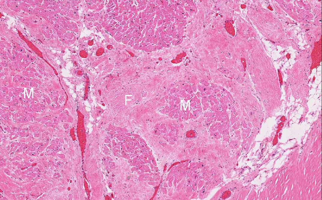
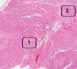
Hematoxylin & eosin |
Area 1: Note the difference between the fibrotic part (F) and residual myocardial fibers (M). Occasional entrapped fibers (arrow) are seen within the fibrotic areas. |
|
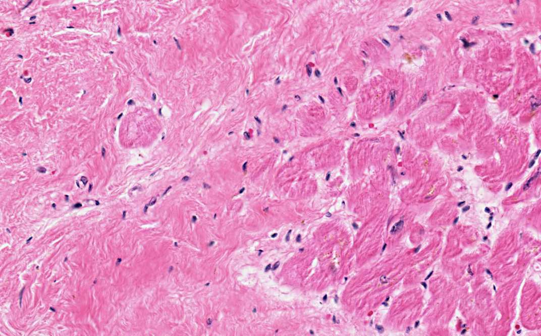
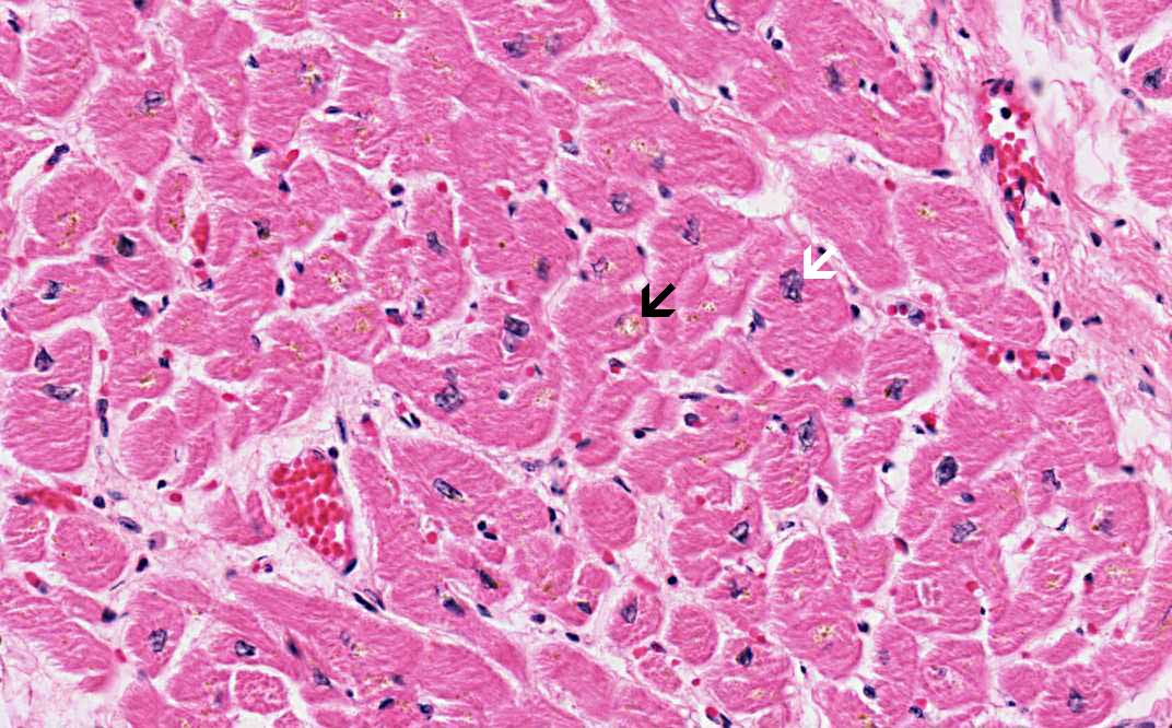
Hematoxylin & eosin |
Area 2: Note that fibrosis is absent in this area. It indicates that this area has not been affected by the infarction to the extent that there is necrosis and fibrosis. Note the enlarged nuclei indicative of myocardial hypertrophy (white arrow). The yellowish depositions (black arrow) are lipofusin. |
|
History: This slide was obtained from the archive and had no history but the patient has a history of remote heart attack.
Histologic Highlights of this Case:
Compare this with other stages of myocardial infarction: C005, C006 |
Original slide is contributed by Pathology Learning Center, University of Iowa (Iowa Image Collection).