Case No.: G-002
Diagnosis: Mixed germ cell tumor, predominantly seminoma with focal embryonal cell carcinoma and yolk sac tumor
Organ: Testis
Last Updated: 12/21/2011
|
|
|
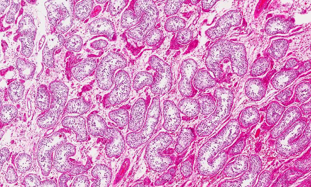
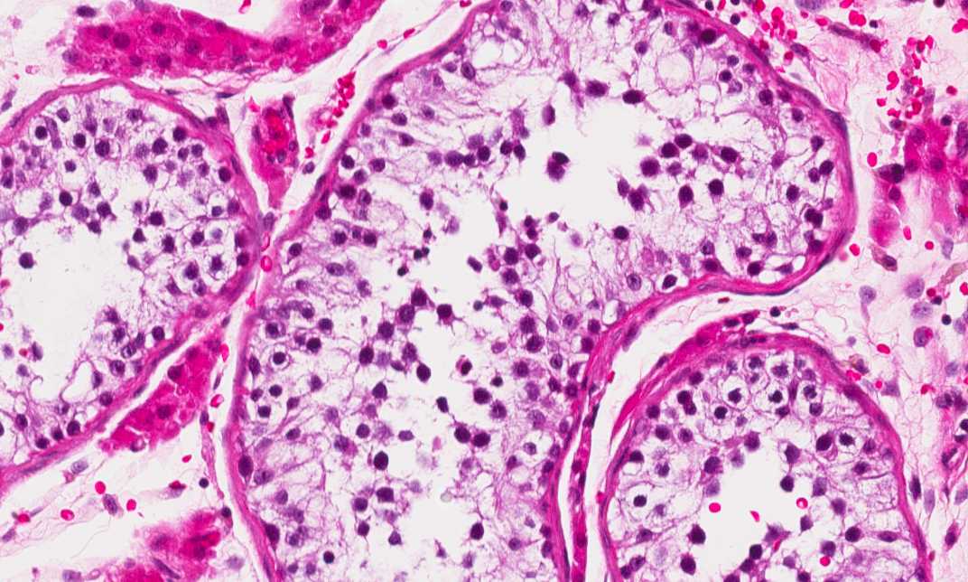
Hematoxylin & eosin |
Area 1: Seminiferous tubules are illustrated here. |
|
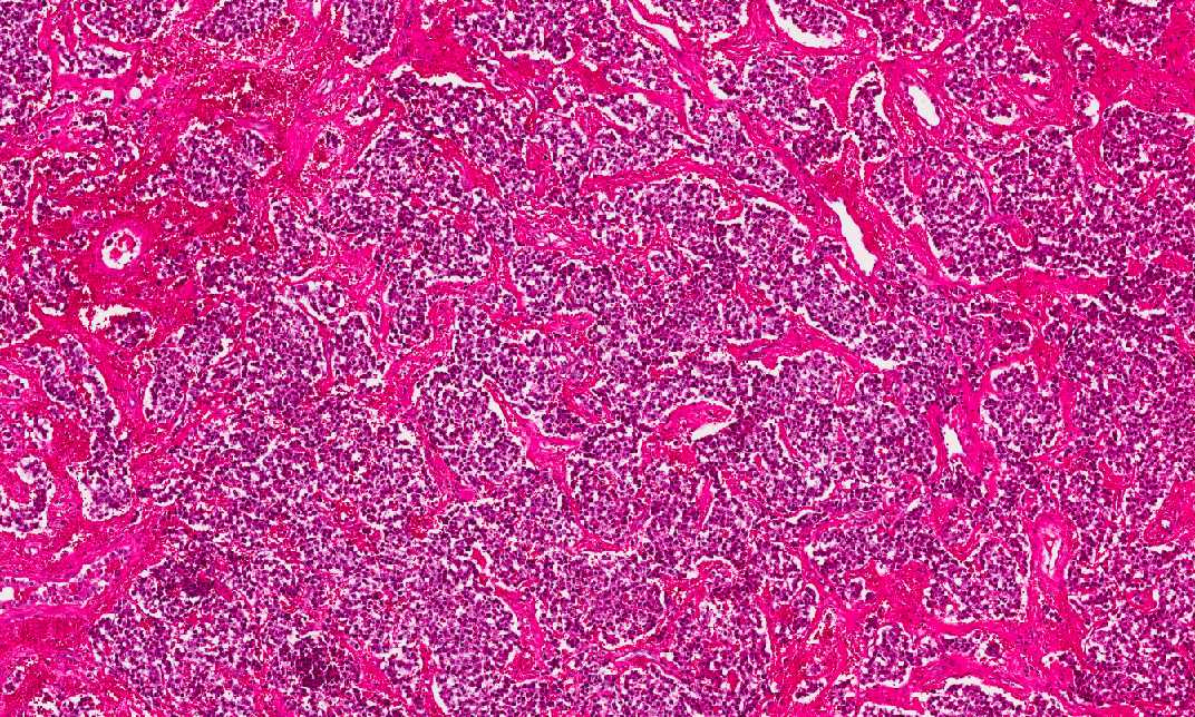
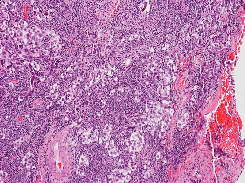
Hematoxylin & eosin |
Area 2: This is the seminomatous component. There is very little lymphocytes accompanying this tumor. See Bonus Images below. |
|
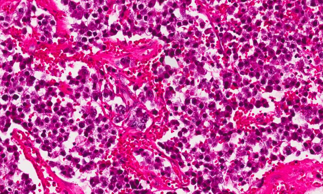
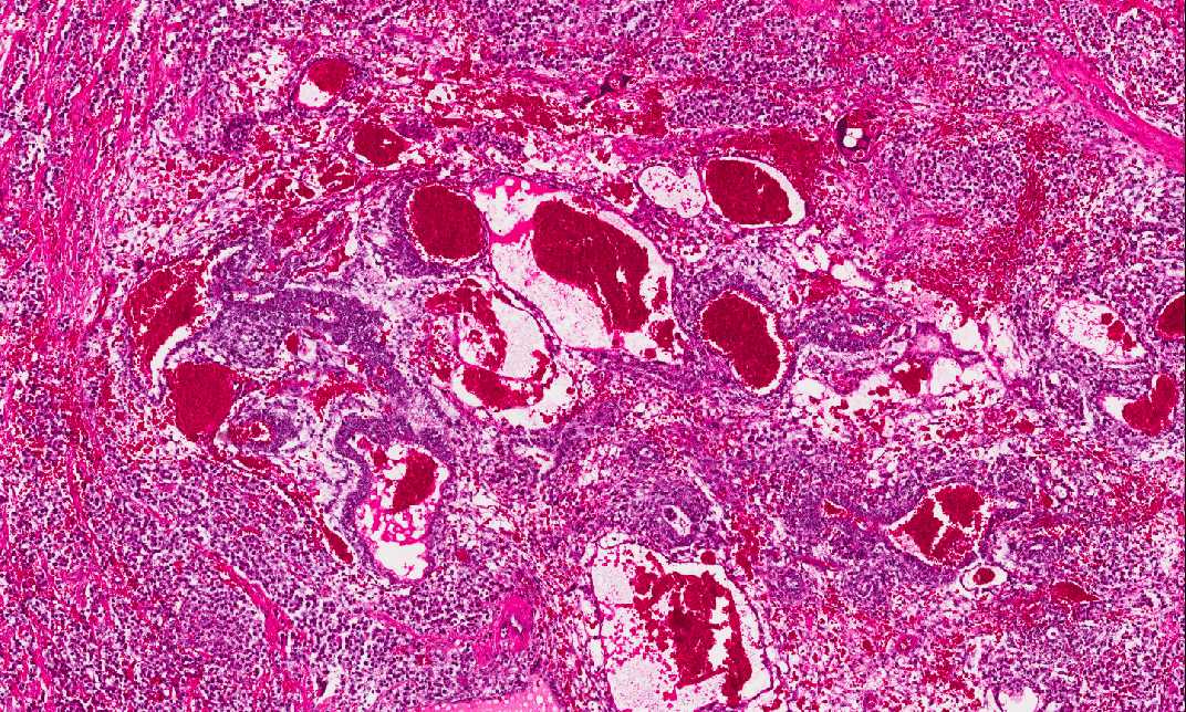
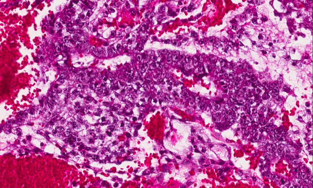
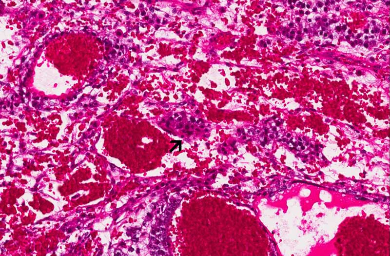
Hematoxylin & eosin |
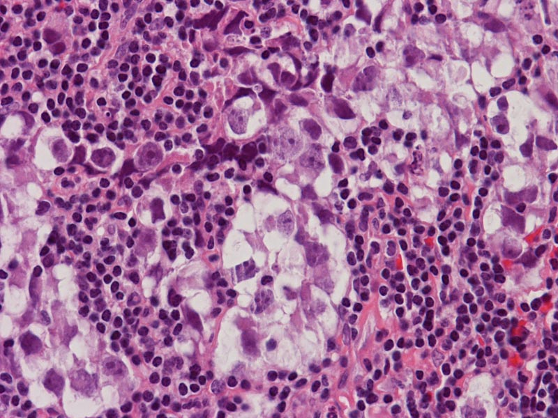
Area 3: This neoplastic foci is composed of tumor cells significantly larger than the seminomatous component. While some of the areas are composed of solid sheets of large pleomorphic cells, there are other areas that arrange into structures with a lumen. These areas represent a mixture of embryonal cell carcinoma and yolk sac tumor. Scant syncytiotrophoblasts are also noted (arrow). |
|
History: This specimen was obtained from a 28 year-old man. What is this organ? What is your diagnosis?
Histologic Highlights of this Case:
Comment:
|
Bonus Images:
|
Hematoxylin & eosin |
Germinoma from pineal gland: These images are obtained from a germinoma of the pineal glands. The fixation is optimal and the morphology of the seminomatous neoplastic cells can be clearly seen. They are usually large round to polygonal, with a large nuclei and prominent nucleoli. Also, a substantial amount of lymphocytes are also present in contrast to our current. |
Original slide is contributed by Dr. Kar-Ming Fung, University of Oklahoma Health Science Center, Oklahoma, U.S.A.