|
History: The patient was a 47
year-old woman who presented to her doctor with the chief complain of
difficulty in breathing and cough. On further work up, a large anterior
mediastinal mass was revealed by CT scan. She also has a history of
uterine cervical carcinoma. The differential diagnoses metastatic
carcinoma, thymoma, lymphoma, ans soft tissue tumors. The current specimen was obtained from the thymectomy which yielded a 14.0 x 15.0 x 4.5 cm mass.
Gross Pathology:
Histologic Highlights of this Case:
-
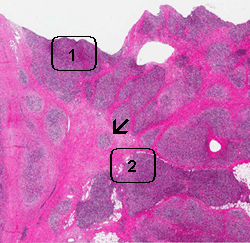
This tumor has several notable features.
It is composed predominantly of small lymphocytes without any
suggestion of lymphoma. Among these lymphocytes are scattered large,
atypical cells with one or, less commonly, multiple nuclei (area 1).
Many of the nuclei are lobulated and typically a prominent,
eosinophilic nucleoli are present. The distribution of these cells
are not homogeneous. They are hard to find in some areas. Also
present in this backgound of lymphocytes are eosinophils (area 2).
Again, their distribution is also non-homogeneous. The background of
this tumor is very fibrotic and many nodules (arrow) surrounded by
the fibrous tissue is present. These features are classic for
nodular sclerosing type of Hodgkin lymphoma. The large atypical
cells in fact are subclassified into the several categories as
discussed below. Many of them
can be found in this case.
Characteristic Cells in Hodgkin Lymphoma:
-
Reed-Sternberg (RS) cells: Classical
diagnostic Reed-Sternberg are large cells with abundant basophilic
cytoplasm and must have at least two nuclear lobes or nuclei. The
nuclei are large and often rounded in contour and have a prominent
eosinophilic nucleolus. Diagnostic RS cells must have
lat least two nucleoli in two separate nuclear lobes. These cells,
however, do not need mirror-image double nuclei to be classified as
Reed-Sternberg cells. Classical
-
Hodgkin cells are uninuclear
version of Reed-Sternberg cells.
-
Lacunar cells are characteristic
of nodular sclerosing classical Hodgkin lymphoma and the
characteristic feature is retraction of the cytoplasm from the
surrounding and thus the cell apparently lie within lacunae. The
cytoplasmic retraction, however, is produced by formalin fixation
because these cells are not seen in specimens fixed in B5 or Zenker
fixatives.
-
Pleomorphic Reed-Sternberg cells
have large bizarre polyploid nuclei.
-
Mummified cells are large
necrotic cells with deep eosinophilic cytoplasm and contracted
contour. The nuclear details are also lost.
-
"Popcorn cells":
These cells have multilobated nuclei and closely
resemble RS cells. Unlike RS
cells, however, the nuclei contain multiple small nucleoli and they
lack CD15 or CD30 immunoreactivity. Rather, they are positive for
CD45 and other B-cell markers (CD20, CD79a). They are typically seen
in nodular lymphocyte predominant Hodgkin lymphoma. This tumor
behaves biologically similar to large B-cell lymphoma. However, the
neoplastic lymphocytes are sparse which grant them histopathologic
similarities to classical Hodgkin lymphoma.
Classification of Hodgkin lymphoma:
-
In the World Health Organization (WHO)
classification, Hodgkin Lymphoma is classified into:
Nodular lymphocyte predominant Hodgkin lymphoma
Classic Hodgkin lymphoma
Nodular sclerosing classical Hodgkin lymphoma
Lymphocyte-rich classical Hodgkin lymphoma
Mixed cellularity classical Hodgkin lymphoma
Lymphocyte-depleted classical Hodgkin lymphoma
-
In order to make a diagnosis of the
nodular sclerosing type, there must be collagen bands that surround
at least one nodules and lacunar type Reed-Sternberg cells.
|
