Case No.: H-004
Diagnosis: Spleen
Organ: Gaucher disease
Last Updated: 12/21/2011
|
|
|
Hematoxylin & eosin |
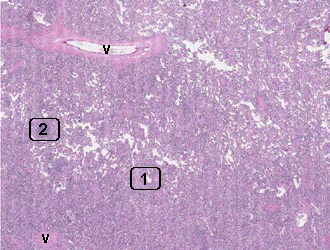
Area 1: The Gaucher cells are present through out the spleen and lead to splenomegaly. Note that many Gaucher cells have a vaguely laminated cytoplsm (arrow). |
|
Hematoxylin & eosin |
Area 2: Only a small amount of white pulp is present (arrows). |
|
History: This slide was obtained from an old teaching set and the history was not provide. However, patient affected by this disorder often have painless hepatomegaly and splenomegaly, hypersplenism and pancytopenia. Many of them would experience severe bone pain with the hip and the knee most affected. Although this disorder is usually adult onset, childhood and juvenile onset can also be seen. Not all of these patients are affected by neurologic manifestations. This condition is most common in Jewish people of Eastern and Central European descent (Ashkenazi Jews).
Histologic Highlights of this Case:
Comment:
|
Bonus Images:
|
Diff Quic |
Gaucher cells from bone marrow: This is a high-magnification of a Gaucher cells obtained from a bone marrow smear and staind with Diff Quick stain. The laminated cytoplasm that are characterized as "crumbled tissue paper" is well highlighted here. |
Original slide is contributed by James Fishback MD, Department of Pathology, Kansas University (Iowa Image Collection).