Case No.: J-001
Diagnosis: Squamous cell carcinoma, non-keratinizing
Organ: Lung, left upper lobe
Last Updated: 12/21/2010
|
|
|
Hematoxylin & eosin |
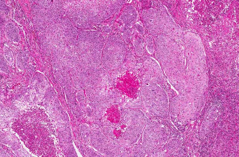
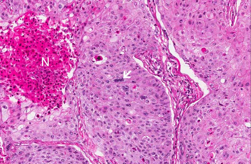
Area 1: The tumor cells form squamous cell nests with central necrosis (N) in some of them. The nuclei of the carcinoma cells are large and with prominent nucleoli. Mitoses are common (arrow). |
|
Hematoxylin & eosin |
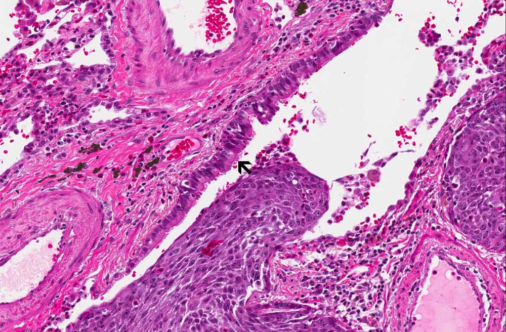
Area 2: This image is taken at the periphery of the tumor. Note that a segment of residual bronchus is still present. Note that the bronchus is lined by ciliated columnar epithelium (arrow). |
|
|
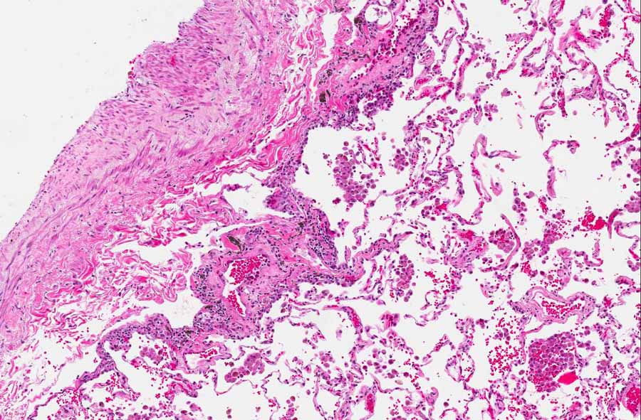
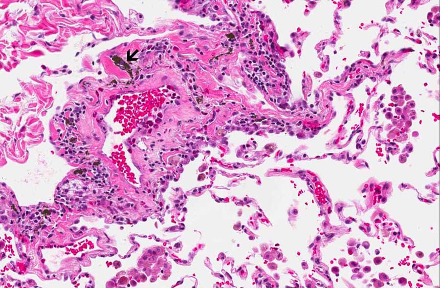
Area 3: This image is taken from the areas uninvolved by the carcinoma. Note the anthracotic pigment depositions (arrow). |
|
History: The patient was an 81 year-old man with a mass in the right upper lobe. The lesion was excised through a lobectomy and yielded the current specimen. On gross examination, the mass was a roughly round to oval nodule, 2.6 cm in greatest dimension. It was well demarcated from the lung parenchyma and did not involve the overlying visceral pleura.
Histologic Highlights of this Case:
Immunohistochemistry:
|
Bonus Images:
|
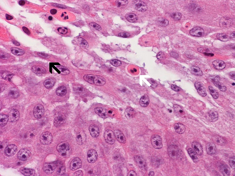
Hematoxylin & eosin |
High magnification image: This image is taken at high magnification. The intercellular bridges (black arrow) is well illustrated. There are also dyskeratotic cells (white arrow). Both are features of squamous cell carcinoma. There is, however, no true keratin pearl formation in this case. Note the large nuclei and their prominent nucleoli. |
|
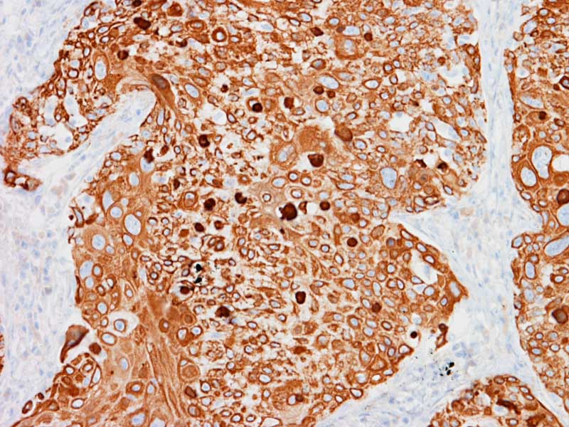
Cytokeratin 5/6 |
Cytokeratin 5/6: Cytokeratin 5/6 is often expressed in squamous cell carcinoma but not adenocarcinoma. It offers a way to differentiate the two in difficult cases. |
|
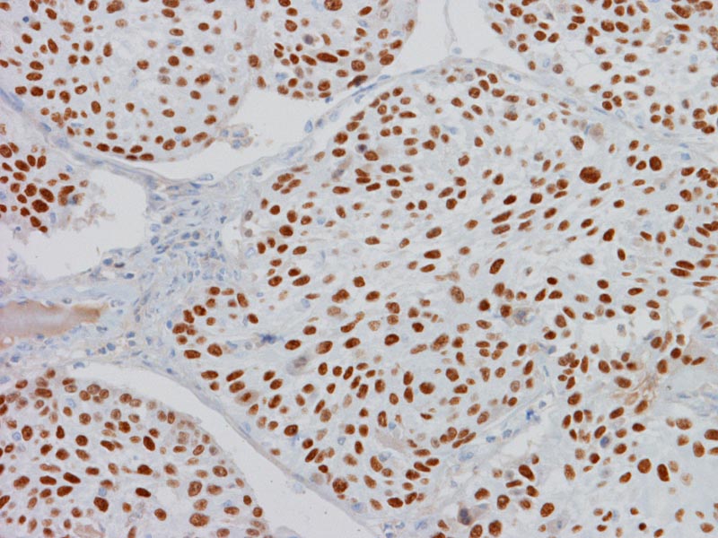
P63 |
P63: P63 is often expressed in squamous cell carcinoma but not adenocarcinoma. It offers a way to differentiate the two in difficult cases. |
Original slide is contributed by Dr. Kar-Ming Fung, University of Oklahoma Health Sciences Center, Oklahoma, U.S.A.