Case No.: J-002
Diagnosis: Squamous cell carcinoma, keratinizing
Organ: Lung, right upper lobe
Last Updated:
12/21/2010

Online Slide/Full
Screen/
Open with ImageScope
|
|


Hematoxylin & eosin |
Area
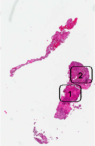
1: Note the keratinizing pearls (arrow in the low-magnification
image) and the intecellular bridges (arrow in the high-magnification
image. |
|


Hematoxylin & eosin |
Area
2: Note that other areas are involved by the tumor but, in addition,
there are also foamy macrophages (arrow) |
|
|
History: A 65 year-old woman
presented with a lung mass in the right upper lobe. An endobronchial
biopsy was performed and yielded the current specimen.
Histologic Highlights of this Case:
-
This is a small endobronchial biopsy.
The salient features are the keratin pearls (area 1) which is a
diagnostic feature of squamous cell carcinoma. Intercellular bridges
are also present which is also a classic feature of squmous cell
carcinoma.
-
In some areas of the the specimen, some
foamy cells are present and are likely to represent foamy
macrophages. They are rather common in lung alveoli distal to
obstruction of the corresponding bronchus. As lung cancer often
generates this kind of obstruction, foamy histiocytes are rather
common. (area 2)
-
Click here to see a squamous cell carcinoma of lung without keratin
pear formation.
|
Original slide is contributed by Dr. Kar-Ming
Fung, University of Oklahoma Health Sciences Center, Oklahoma, U.S.A.
Home Page


