Hematoxylin-Eosin
Luxol fast blue-PAS
Case No.: N-011
Diagnosis: Demyelinating disease
Organ: Cerebral hemisphere
Last Updated: 1/31/2010
|
Hematoxylin-Eosin |
Luxol fast blue-PAS |
|
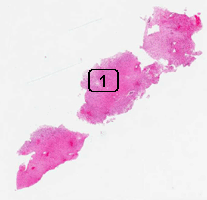
Hematoxylin & eosin |
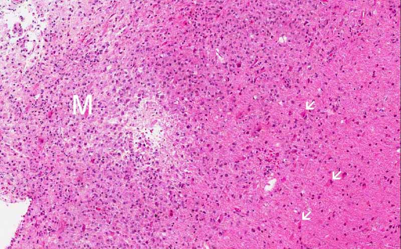
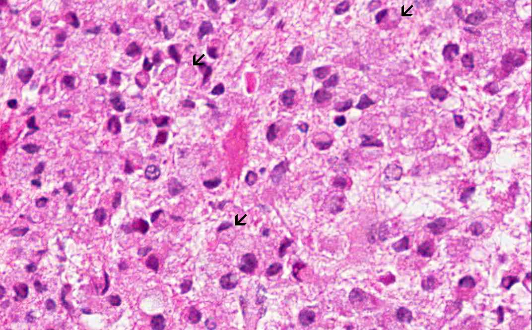
Area 1: This image is taken at the interface of the area extensively infiltrated by macrophages (M) and area far less significantly infiltrated by foamy macrophages and many reactive astrocytes (white arrows) are present in this area. On high magnification, note that the foamy histiocytes have a moderate amount of cytoplasm with a bubbly or foamy appearance. Hence they are called foamy macrophages. |
|
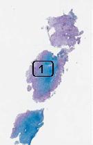
Luxol fast blue & PAS |
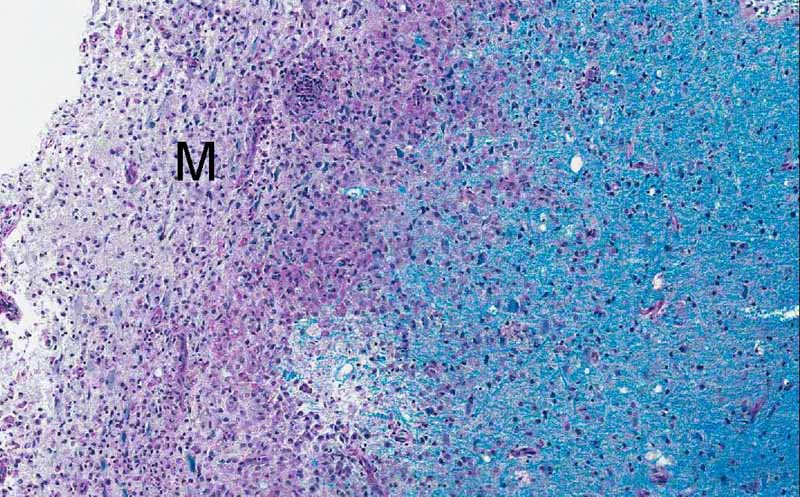
Luxol Fast Blue and PAS Stain: Luxol fast blue demonstrates normal myelin in blue. This photo is taken from area 1 and you can see that the area infiltrated by macrophages does not have much myelin but the area that is not infiltrated by macrophages has myelin. This indicates a lost of myelin. This is a classic histopathologic finding. The macrophages are scavengers that remove the degraded myelin. The PAS stain is a counter stain but it has the ability to stain up the lysosomes in macrophages red. |
|
Immunohistochemistry |
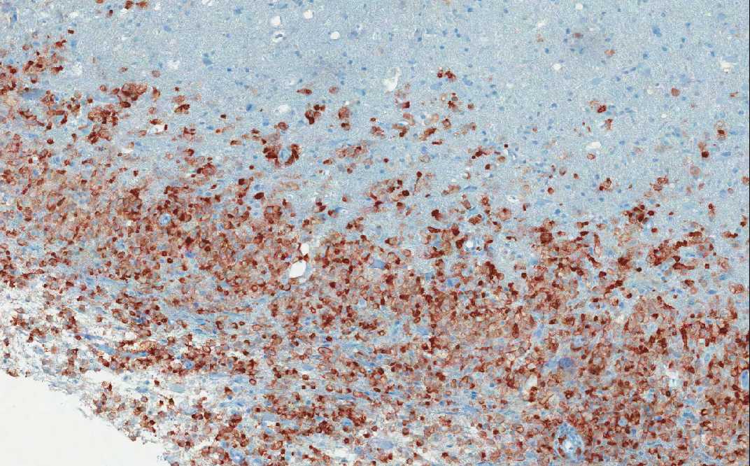
CD163: This is a marker for macrophages and the immunohistochemistry highlights the macrophages (brown cells). This photo is also taken from area 1 and you can see the well demarcated macrophages from the white matter with preserved myelin. Note that there are also some macrophages infiltrating into the white matter where myelin is still present but the number is small. It may be difficult to identify these cells based on morphologic features alone and immunohisochemistry for CD163 is a big help in identifying these cells. |
|
Immunohistochemistry |
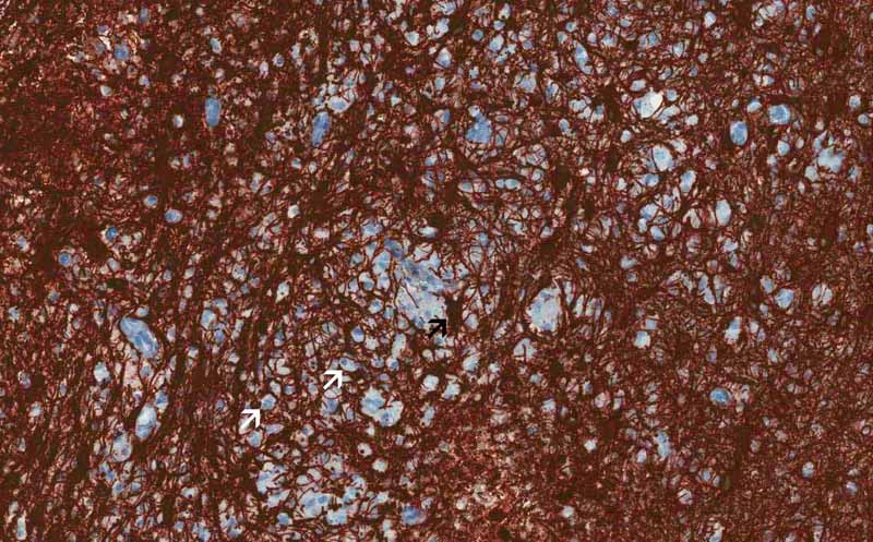
Neurofilament proteins: Neurofilament proteins are intermediate filaments. A large amount of them are found in the axons and therefore immunohistochemistry for neurofilament proteins would allow us to demonstrate the proteins. Some silver based stains such as Bodian stain is also appropriate for this purpose. This photo is taken from the same area similar to the one used in CD163. You can see foamy macrophages (arrows) on one side of the image but not the other side. In between the macrophages are axons that are positive for neurofilament proteins. Compare this with the Luxol Fast Blue and PAS stained section, you can see that there is tremendous loss of myelin in area infiltrated by macrophages but the axons are preserved. This is a key feature of demyelinating disease. |
|
Immunohistochemistry |
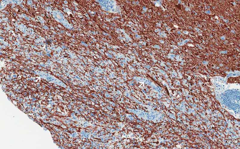
Glial fibrillary acidic protein (GFAP): GFAP is also an intermediate filament and is found in astrocytes. The image here demonstrate a dense mesh of GFAP cytoplasmic processes with foamy macrophages (white arrows) scattered in between. Astrocytes has star like processes which makes them look like a star and hence their name (black arrow). Click here to see another image of stellate shaped reactive astroytes stained for GFAP. This dense mesh of astrocytes is abnormal and is resulted from proliferation of reactive astrocytes due to the demyelinating process. |
|
CD68 |
Neurofilament |
GFAP |
|
History: The patient was a 38 year-old woman with complain of headache for 3 months. MRI showed multiple lesions in both cerebral hemispheres with some of the lesions touching the ventricles. The patient was HIV (-) and not immunosuppressed. A stereotactic biopsy was performed and yielded this specimen.
Histologic Highlights of this Case:
|
Original slide is contributed by Dr. Kar-Ming Fung, University of Oklahoma Health Sciences Center, Oklahoma, U.S.A.