Case No.: N-019 Quiz

Diagnosis: Diffuse astrocytoma, WHO grade II

Organ: Brain, frontal-temporal region

Last Updated: 12/21/2010
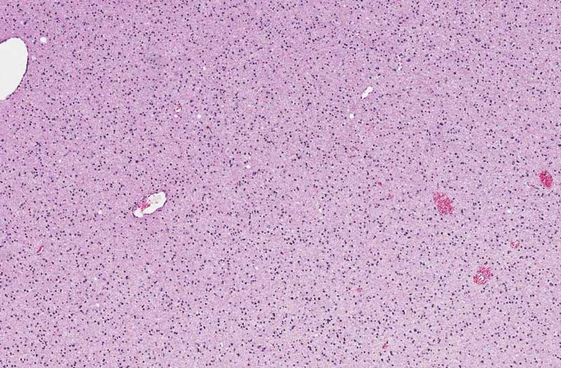
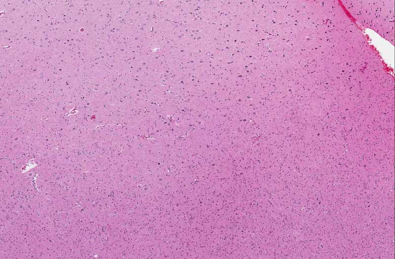
 

Online Slide/Full Screen/Open with ImageScope

Hematoxylin & eosin

Area 1: The cellularity (the number of cells per unit area) in the deep white matter is increased. The cytoplasmic processes are rather coarse and in between the processes are some bluish mucoid material. These material are very classic for low grade glial neoplasm. Microcysts are often a common feature of low grade glial neoplasms although they are not present here. These cysts often contain this type of mucoid material.
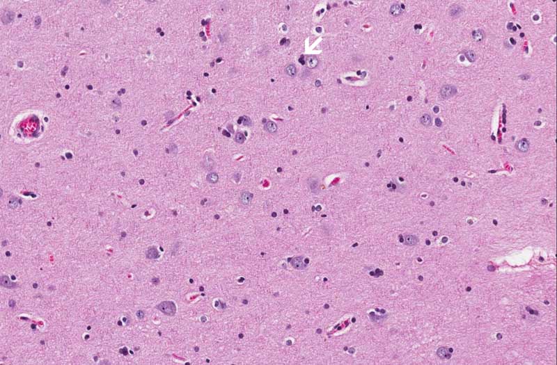
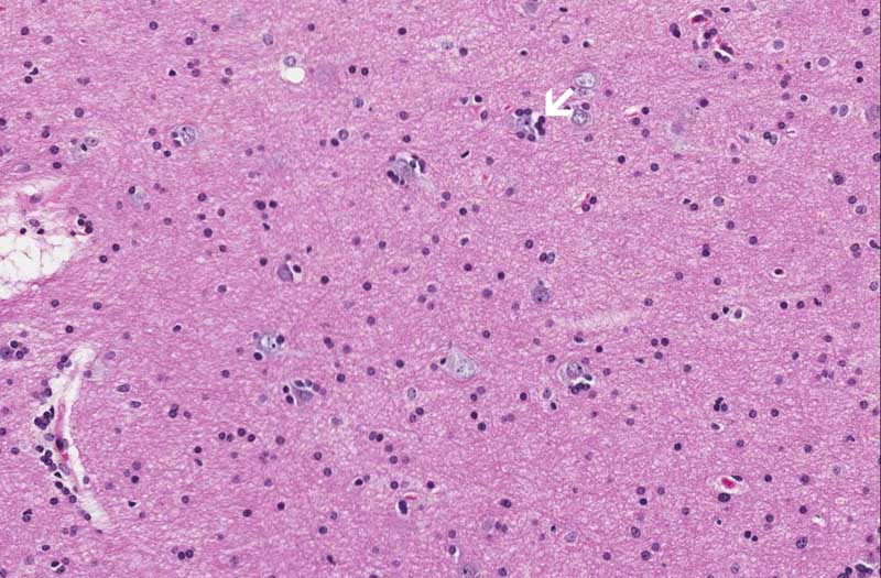
 

Hematoxylin & eosin

Area 2: These images are taken from an area where the cortex is infiltrated by the tumor. Image B is taken from a place more superficial (farther from the tumor) than image C which is taken from a location closer to the tumor. Note the tumor cells circling neurons (arrow) and this phenomenon is known as satellitosis (arrows in  B and C).
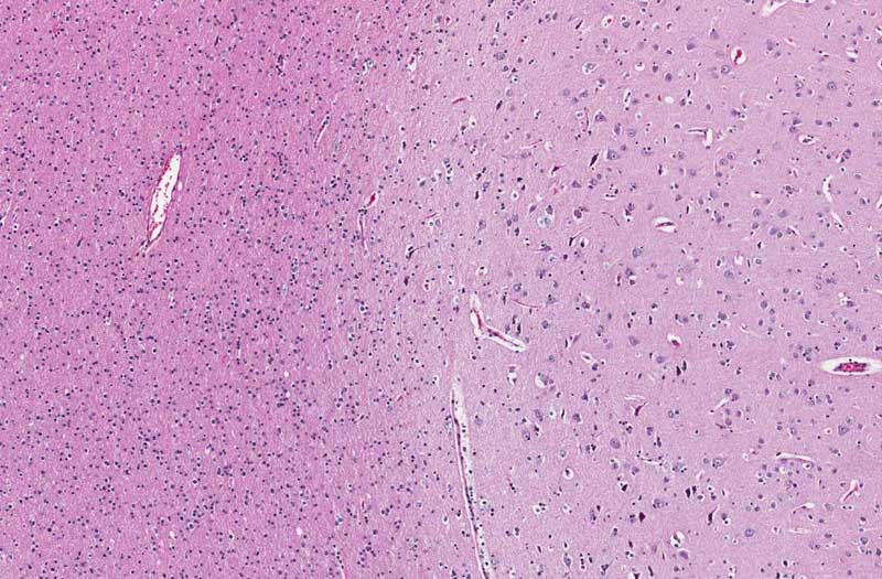
Hematoxylin & eosin

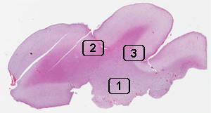
Area 3: There is still increased in cellularity in the white matter indicating the presence of tumor in this area. However, the gray-white junction is much more distinct in comparison to area 2.

History: The patient was a 25 year old woman who complained of headache. MRI revealed a large, non-enhancing tumor in the frontal-temporal lobe. The mass was excised and yielded the current specimen.


Histologic Highlights of this Case:

  • The general configuration of the cortex and subcortical white matter is preserved in this specimen. However, the deeper part of the white matter is replaced by a cellular neoplasm (area 1). Note that practically all of the tumor cells have small nuclei and minimal pleomorphism. On a single cell basis (cytologic basis), it is not exactly easy to tell the difference between these cells and normal glial cells. Mitotic figures are not readily seen. These features are classic for a diffuse astrocytoma of WHO grade II.

  • Note that the tumor cells infiltrate into the overlying cortex (area 2) and such infiltration. The tumor cells have a tendency to wrap around neurons. If you think the neuron is a planet and then the surrounding tumor cells are then satellites of the planet and hence this phenomenon is known as satellitosis. This phenomen is not limited to astrocytoma. It is a commonly seen feature in oligodendrogliomas too. Satellitosis, however, can also occur in pathologic brain parenchymal tissue without involvment by glial neoplasms. In addition, a light degree of satellitosis may be seen in normal brain parenchyma.

  • In some of the area, the gray matter is less involved and the gray white junction is more distinct (area 3).

Immunohistochemistry:

  • p53: Immunohistochemistry positive for p53 can be seen in a subset of astrocytoma. In tumors that are positive, it is a useful tool in identifying single infiltrating glioima cells particularly at the infiltrating edge of the tumor. In the current case, the morphologic diagnosis is obvious, demonstration of p53 in tumor cells is not particularly helpful in making the diagnosis.

  • Glial fibrillary acidic protein (GFAP): Neoplastic astrocyres are positive for GFAP. In the current case, the differentiation is well enough for a morphologic diagnosis and immunohistochemistry for GFAP would not provide more useful information or help in diagnosis. However, GFAP and oligo2 can be useful markers in confirming the glial phenotypes of some poorly differentiated tumors.

Comment:

  • This specimen is a wonderful demonstration on the diffuse nature of diffuse astrocytoma. The surgical margins of these tumor are basically impossible to be determined with confidence.

Bonus Images:

Hematoxylin & eosin

Luxol fast blue-

Cresyl Violet

For Comparison: This is a section taken from the parietal lobe of a patient (autopsy) with no significant histologic abnormalities. You can compare the cellularity of the white matter and the sharp gray white matter distinction with the current slide.

Click here to see more neuroanatomy slides

Original slide is contributed by Dr. Kar-Ming Fung, University of Oklahoma Health Sciences Center, Oklahoma, U.S.A.

 Home Page