|
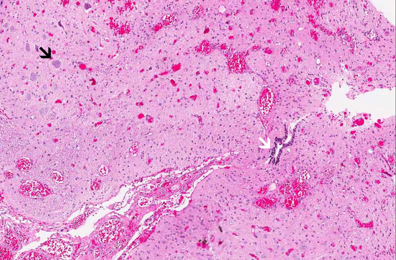
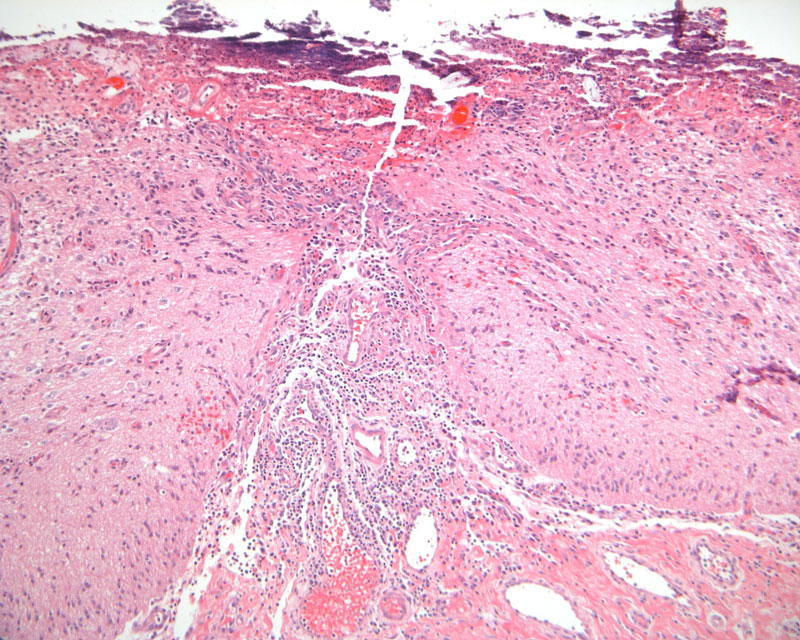
Hematoxylin & eosin |
Area 1: A cleft like space lined by ependymal cells is present in this area (white arrow). The shape of this structure is suggestive of a fragment of the central canal. Some large neurons (black arrow) are also present. These structures, however, are disorganized and this is typical for meningomyelocele. |
|
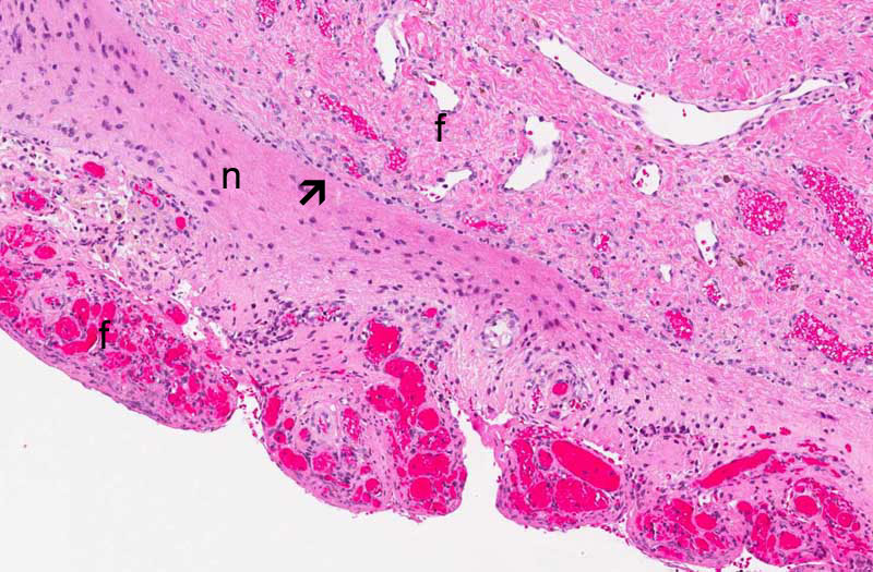
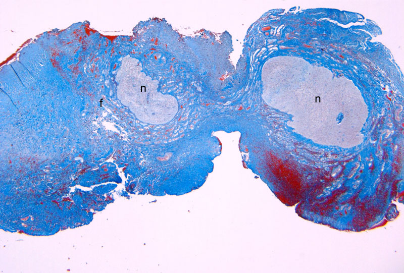
Hematoxylin & eosin |
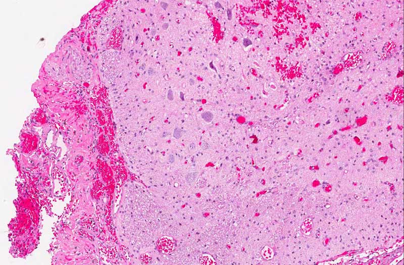
Area 2: There is a rich network of blood vessels attached to the brain parenchyma. The leptomeninges is not readily seen . The border between the blood vessel and the neural parenchyma is well demarcated (arrow). In this particular area, an island of neural parenchymal tissue (n) is trapped in between two portion of fibroconnective tissue with rich vasculature (f). |
|
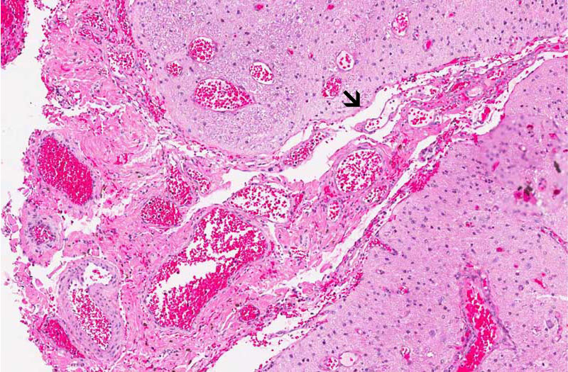
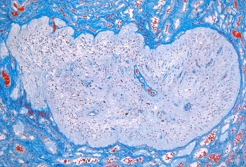
Hematoxylin & eosin |
Area 3: In contrast to Area 2, the blood vessels are not firmly adhered to the brain parenchyma and the leoptmeninges (arrow) can be recognized. |
 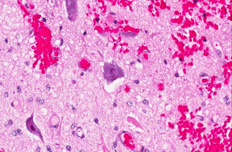 Hematoxylin & eosin |
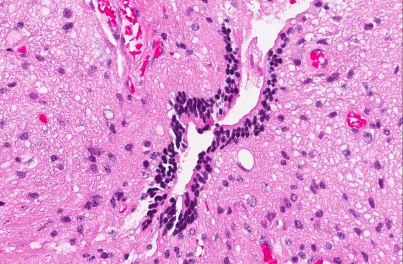
Area 4: Large neurons like the one being illustrated here are present in different part of the specimen. Can you find them? These neurons are large enough to compare with those in the anterior horn cells. This morphologic similarities, however, are not enough proof that these are anterior horn cells. |