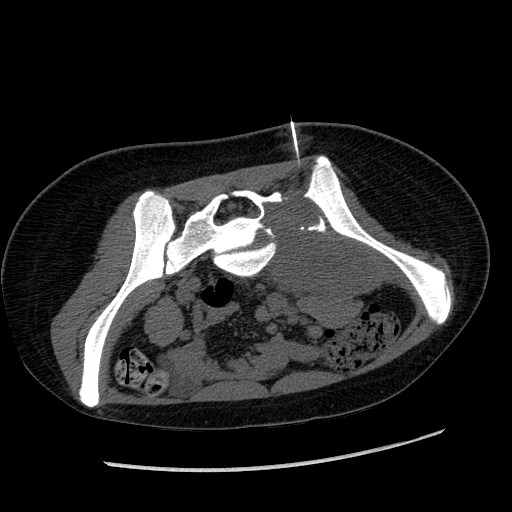
needle biopsy
(the needle is
shown here)
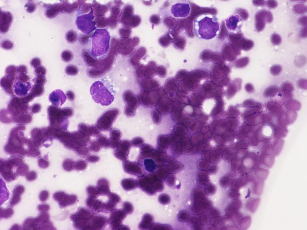
Diff Quick Stain
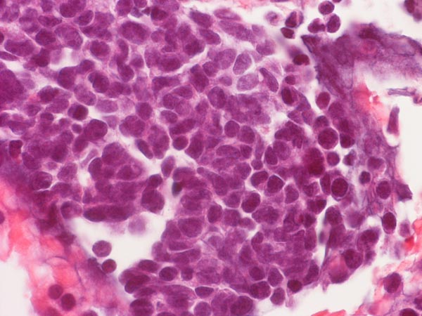
Hematoxylin &
Eosin Stain
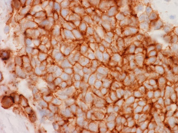
for CD99
Answer and Discussion of Quiz Set: N-032
1. The patient was a 3 year-old girl with the chief complain of left lower extremity that extended to the right side. Imaging demonstrated a large sacral mass that mainly affects her left side. These features are not sufficient for a definitive diagnosis but which of the following be the most likely diagnosis?
| CT guided needle biopsy (the needle is shown here) |
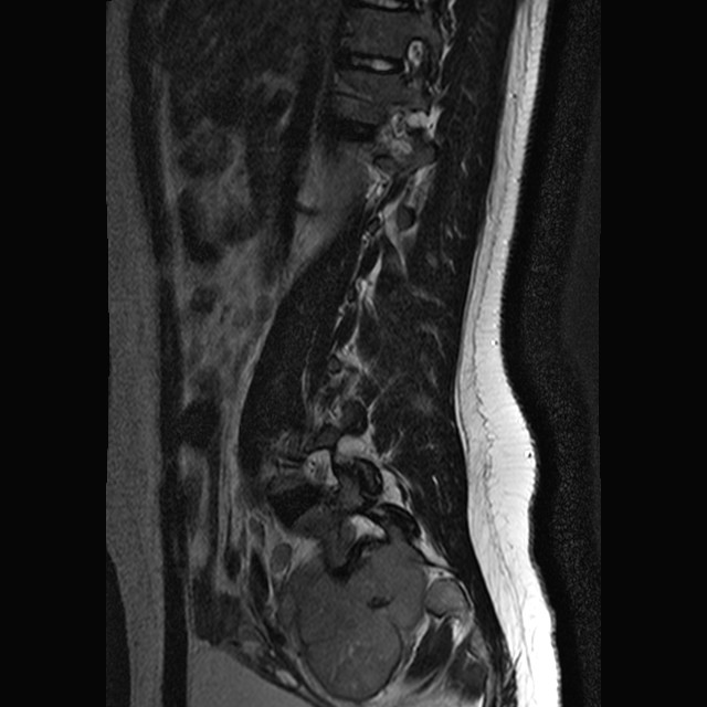
MRI | FNA Diff Quick Stain |
Permanent
section Hematoxylin & Eosin Stain |
Immunohistochemistry for CD99 |
|
|
|
|
|
|
A. Osteosarcoma
B. Ependymoma, WHO grade II
C. Atypical teratoid/rhabdoid tumor (AT/RT)
D. Ewing's sarcoma
E. Metastatic Merkel cell carcinoma
Answer and Discussion: The answer is (E). Although the features initially provided were insufficient for a definitive diagnosis, there are features here that would strongly favor an Ewing's sarcoma. There is a large tumor with bone destruction in the left pelvis that protrudes into the pelvic cavity. On cytologic smear, Diff Quick stain demonstrates neoplastic cells with large nuclei and high nuclear to cytoplasmic ratio. Some cytoplasmic bubbles (arrows) are also present. Histologically, the tumor is that of a small blue cell tumor and the nuclear features are suggestive of an neuroendocrine tumor. This tumor was positive for CD99. On further study with fluorescent hybridization, translocation specific for Ewing's sarcoma was demonstrated.
Osteosarcoma is common in the pediatric age group but they are more likely to be seen in older children and adolescents. In addition, they are more common in long bone than in the axial skeleton in this age group. Osteosarcoma arising from the axial skeleton is more common in older adults. The histology is that of a small blue cell tumor suggestive of a neuroendocrine tumor. This type of histology would be quite unusual for osteosarcoma. Also, there is no neoplastic osteoid formation in the provided image to suggest osteosarcoma.
Ependymoma is a WHO grade II tumor. It is not an uncommon tumor in the spinal cord. However, ependymoma rare invades surround bone and protrude out as a large mass as in this case. Also, there is no histologic features such as perivascular pseudorosettes in the histology to suggest ependymoma. The nuclei of ependymoma usually look lower histologic grade and often has a small nucleoli although anaplastic ependymoma can have enlarged and hyperchromic nuclei. These features are not in favor of an ependymoma.
Atypical teratoid/rhabdoid tumor can occur in this location. It is not common for them to protrude from the spinal canal and form a large mass but that can happen. The Diff Quick smear does not show any enlarged nuclei and the histologic sections does not show any features of rhabdoid changes. If this is an AT/RT, it would be one with rather uncommon histology. One can argue that AT/RT often contains small cell component and that the FNA has targeted the small cell component. In general, the clinical location and the total lack of prominent nuclei and rhabdoid features makes AT/RT not the most likely diagnosis.
Metastatic Merkel cell carcinoma would appear as a neuroendocrine tumor. There is no histologic evidence to go against the diagnosis of metastatic Merkel cell carcinoma. However, the age of the patient is too young of Merkel cell carcinoma. In addition, there is no tumor found on the skin to suggest a primary Merkel cell carcinoma.
Ewing's sarcoma can occur in places other than the long bones and it is a common tumor in the pediatric age group. The cytoplasmic bubbles (arrows) are clue to this diagnosis. These vacuoles are formed when the glycogen content of the Ewing's sarcoma has been partially washed out and is best seen on Diff Quick cytologic preparations. The histologic features of this tumor is that of a small blue cell tumor and is compatible with an Ewing's sarcoma. Therefore, without further study with immunohistochemistry, the most likely diagnosis of this case is an Ewing's sarcoma. This tumor turned out to be CD99 positive and with translocation of an Ewing;s sarcoma.