Case No.: W-004

Diagnosis: Metastatic endometrial sarcoma

Organ: Vertebra, T3

Last Updated: 12/21/2010

Online Slide/Full Screen/Open with ImageScope

Hematoxylin & eosin

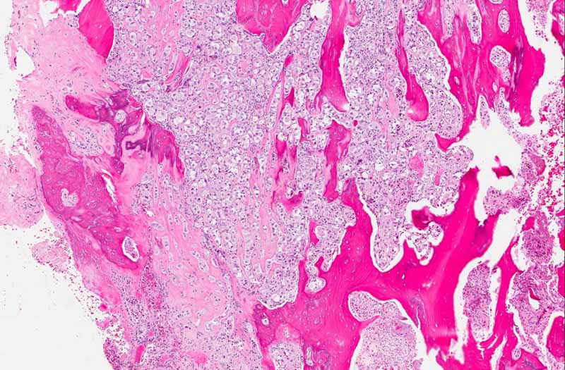
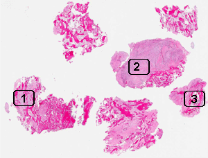
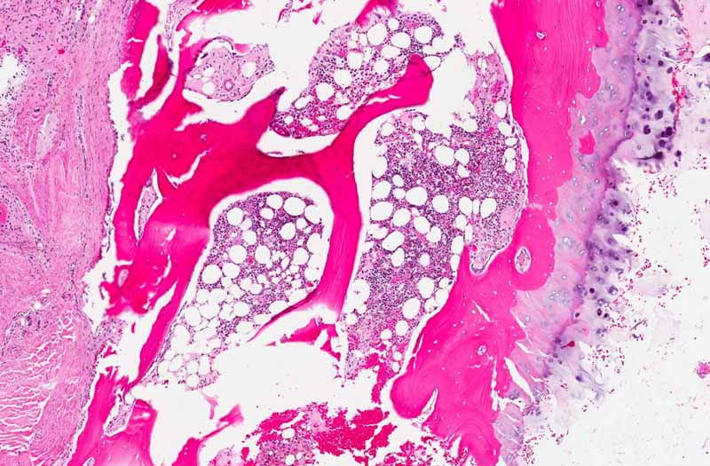
Area 1: The epithelioid features are best preserved in these areas with no or minimal fibrosis. Note the infiltration of the tumor in between the bone fragments.
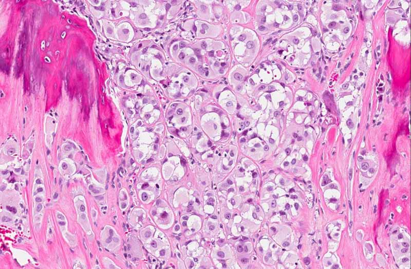
Area 2: In the more fibrotic area, the cells are distorted by the fibrosis and the epithelioid features are seen only focally.
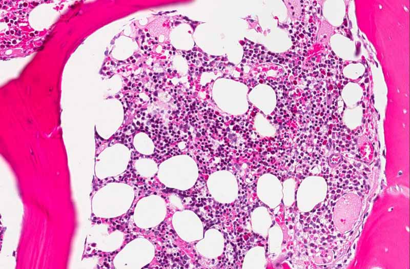
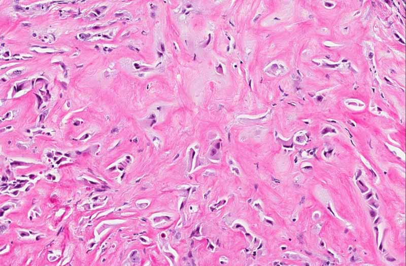
Area 3: This is the area with residual normal bone marrow. Compare the morphology of these cells with the tumor cells.

History: The patient was a 52 year-old woman who was admitted to the emergency room because of back pain. An osteolytic lesion with compression fracture and compression of the spinal cord at vertebral level T3 was uncovered on imaging studies. The lesion was located in the body. As per the patient, she had some kind of gynecologic malignancy and was treated a few years before the current incident. A corpectomy with removal of tumor was performed and yielded the current specimen.


Additional Information: The original pathology report was obtained later and showed that the patient had a history of endometrial sarcoma.


Histologic Highlights of this Case:

  • The bone is extensively infiltrated by large epithelioid cells featured by round to oval cells with a eccentrically located large nuclei. Many but not all of them have prominent nucleoli. No real gland formation or mucinproduction are present. These morphological features are best preserved in areas with no fibrosis (area 1).

  • In areas with dense fibrosis, the shape of the cells are distinctly different. They are rather irregular. The nuclei in these areas do not appear so big and the cells seem to be compressed by the surrounding fibrosis. (area 2)

  • Some residual normal bone marrow is present (area 3). Compare these cells with those of area 1 and 2.

Immunohistochemistry:

  • CD10: Positive in practically all tumor cells.

  • Progesterone receptor: Positive in some tumor cells.

  • Estrogen receptor: Negative in tumor cells.

  • Cytokeratin AE1/AE3, cytokeratin 7, cytokeratin 20, cytokeratin 8/18: Negative in tumor cells.

  • S100: Negative in tumor cells.

  • Vimentin: Positive in practically all tumor cells.

  • Desmin: Negative in tumor cells.

  • Smooth muscle actin: Negative in tumor cells.

Comment:

  • Immunohistochemistry:  The epithelioid morphology raises the suspicion of a metastatic carcinoma particularly when there is a know history of gynecologic malignancy. However, these cells do not have the classic appearance of metastatic carcinoma cells. On the other hand, this type of epithelioid cells can be seen in a variety of sarcoma which includes endometrial sarcoma (cytokeratin negative, CD10 positive). immunohistochemistry for cytokeratins will be an important step to rule in or rule out metastatic carcinoma in this case. The immunohistochemistry does not show any marker that support the idea of metastatic carcinoma. In contrast, the positive immunoreactivities for CD10 and progesterone are support of a diagnosis of metastatic endometrial sarcoma. The know history of endometrial sarcoma is also very helpful.

Original slide is contributed by Dr. Kar-Ming Fung, University of Oklahoma Health Sciences Center, Oklahoma, U.S.A.

 Home Page