Calponin
EMA
PanCK
| A 45 year-old Man with a Submandibular Mass. April, 2003, Case 304-3. Home Page |
Cheng Z. Liu, M.D., Ph.D. and Kar-Ming Fung, M.D., Ph.D. Last update: April 30, 2003.
Department of Pathology, University of Oklahoma Health Sciences Center, Oklahoma City, Oklahoma
Clinical information: 45 year-old man with a mass in left submandibular gland.
Pathology of the case:
Gross pathology: Surgery yielded a 2 cm partly well demarcated and partly poorly demarked, slightly irregular fibrous non-necrotic mass involving the submandibular gland.
Histopathology:
|
|
|
|
|
|
|
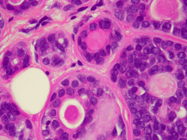
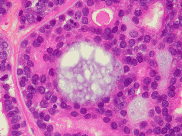
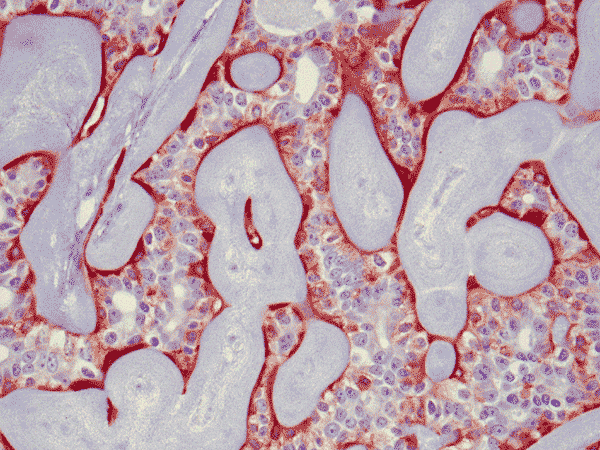
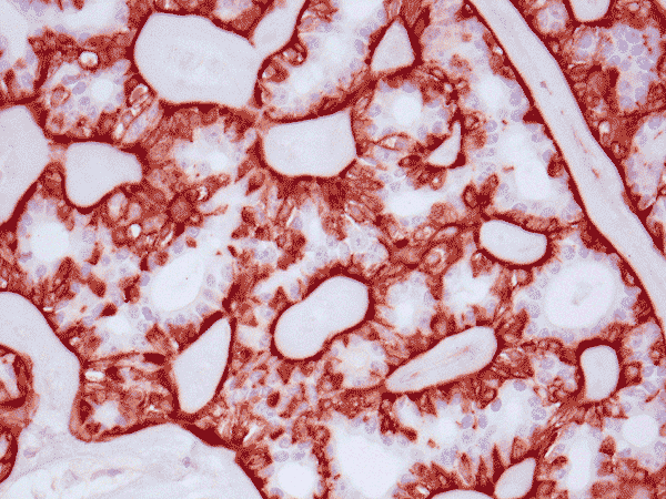
| A. | B. | C. | D. | E. | F. |
|
|
|
|
|||
|
G. Calponin |
H. EMA |
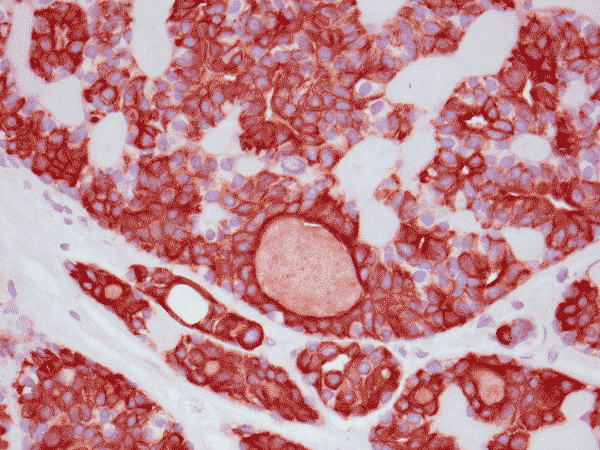
I. PanCK |
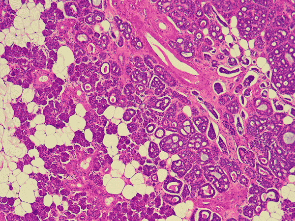
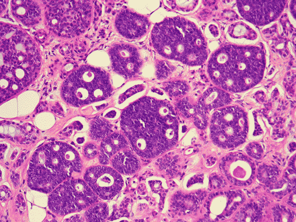
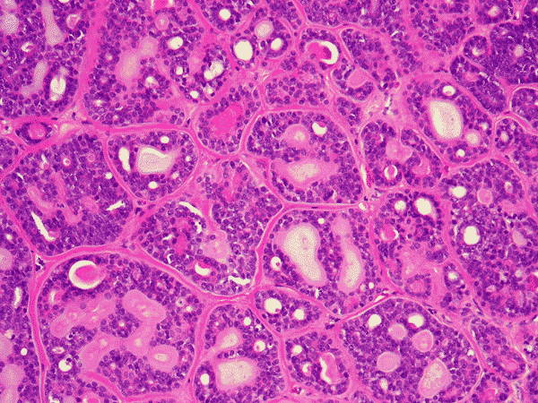
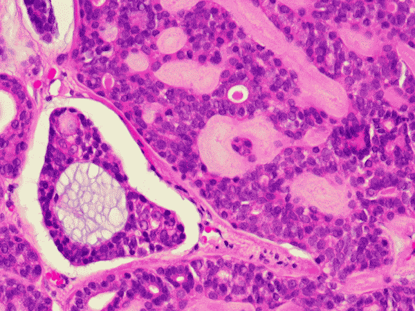
Panel A is a low-magnification photo taken at the edge of the tumor that shows tumor invasion into the surrounding salivary gland tissue. Well formed cribiform arrangment by tumor cells are show in panel B, C, and D; a fibrous stroma can be well appreciated in these photographs. Eosinophilic basement membrane like material can be well recognized in panel E; mucoid material containing microcysts are well illustrated in panel D and F. The myoepithelial component are recognized by strong immunoreactivity for calponin and smooth muscle actin (SMA) in panel G an H; note that the luminal epithelial cells do not have such immunoreactivity. Strong immunoreactivity of the tumor cells for pancytokeratin are well demonstrated in panel I.
| DIAGNOSIS: Adenoid cystic carcinoma, low-grade, of the submandibular gland. |
Discussion: General Information Pathology Differential diagnosis
General Information
The
first histopathologic description of adenoid cystic carcinoma was in 1853 by Robin and Laboulbene.
They used the term tumeur heteradenique, suggesting that it was a
glandular neoplasm arising in a nonglandular area. Billroth employed the term zylindrome
(cylindroma) in 1859. It had certain merits in terms of morphologic implication
but brought confusion with the benign dermal eccrine tumor of the same name. The
term adenoid cystic carcinoma (ADCC) was popularized by Foote and Frazell in
1953 and is preferred term.
Adenoid
cystic carcinomas (ADCC) are slowly metastasizing but relentlessly growing
tumors that often lead to fatality because of local invasion. ADCC makes up
about 10-15% of all salivary gland tumors and have a predilection for the minor
salivary glands. ADCC
are most commonly seen in the 5th
to 7th decade. They are more common in the minor than major salivary
glands and they are one of the most common tumors in the minor salivary glands;
the most common sites are the palate, maxillary sinus, base of tongue, larynx
and trachea. About slightly less than half of the ADCC occurring in the major
salivary glands are found in the submandibular gland
1.
ADCC most likely arise from neoplastic transformation of reserve cells of the terminal duct system. The tumor cells appear to be able to differentiate along the lines of ductal or myoepithelial cells. This supported by the presence of epithelial-type of mucin in the true ducts and basement membrane-like material in the pseudocysts. Immunohistochemistry, in addition, supports the presence of both ductal and myoepithelial cells in these tumors as shown above. The clinical stage is the most important prognostic parameters. Histological grade also correlate with prognosis. Grade I, II, and III have cumulative 15-year survival rates of 39, 26, and 5%. Tumors arising from the sinunasal regions tend to present at higher clinical stages; tumors arising from the submandibular gland tend to be more aggressive. ADCC tends to metastasize to the lung, liver, and soft tissue; metastases to the lymph nodes are infrequent.
The diagnosis of ADCC
can be quite difficult because of the diversified histologic pattern. Correct
diagnosis is very important since many tumors closely mimic ADCC. Recognition
of the two-cell type arrangement and basement membrane depositions are the key
features for correct diagnosis.
The histologic features are well illustrated in this case. In general, ADCC
is a malignant tumor that is clearly invasive at macroscopic and microscopic
levels. Grossly, they are usually fibrous mass with invasion into the
surrounding tissue. The stroma of ADCC is typically fibrotic. The common
histologic patterns are the cribiform pattern, tubular pattern and solid
pattern. The less common pattern are the spindle cell pattern, trabecular
pattern and solid type with comedo-type necrosis. Common
histologic features are cribriform or microcystic architecture with relatively
uniform cells, with small, hyperchromatic nuclei, scant cytoplasm, somewhat
nulcear molding and indistinct cytoplasmic border. These cells are considered as
myoepithelial differentiation.
A second cell type can be recognized as somewhat larger, with more
abundant cytoplasm and less dense nuclear chromatin. These cells are thought to
represent ductal epithelial cells. The ductal cells give rise to real tubular
structures that contain bluish mucoid material; these cells are immunoreactive
for carcinoembryonic antigen (CEA), epithelial membrane antige
(EMA), keratin, and S-100 protein. The myoepithelial cells may arrange in
solid sheets and also give rise to microscystic structures containing hyalinized
eosinophilic basement membrane-like material. They bear immunoreactivity for
calponin,
muscle
specific actin, and low molecular weight cytokeratin. They are also
positive for S-100 protein but are usually less strong than the ductal cells in
the same tumor. Many ADCC also express C-kit (CD117)
2, especially in
solid type. PAS (+) basement membrane-like material is seen in the pseudocyts
and mucin in the true ducts. Perineural invasion is the rule. The tumor cells
are highly proliferative and a Ki-67 labeling index over 20% is not uncommon.
Differential diagnosis
In
general, low-grade ADCC have tubular or cribiform pattern with no solid
area and no necrosis; they are histologically bland and have few or no mitotic
figures. Low-grade tumors are usually small, may have a capsule, and are
amenable to complete excision. Medium-grade tumors often have pure cribiform or
mixed pattern; small amount of solid areas (<30%) is allowed. They also have
increased atypia. High-grade ADCC are usually large tumors that are
difficult to be completely excised. They often have necrosis and substantial
amount of solid areas (>30%) together with active mitotic activity and
pleomorphism. Solid type of ADCC is often a diagnostic challenge since they
closely mimic poorly-differentiated squamous carcinoma, basaloid squamous cell
carcinoma, and small cell undifferentiated carcinoma. Sporadic glandular
structures, focal cribiform arrangement, basement membrane-like material, and
identification of two cell types are helpful features to separate ADCC from
other entities. Solid type of ADCC comprise about 10% of all ADCC and are more
frequently seen in young adults and in minor salivary glands.
Basal
cell adenomas can mimic solid type of ADCC but they are non-invasive; a
feature that distinctly separates them from ADCC. Basal cell adenomas are
uncommon (1-2% of all salivary gland tumor). They occur in late adulthood and
about 70% occurs in parotid glands with the remainder occuring in submandibular
and minor salivary glands. The tumors are composed of nests to trabeculae of
neoplastic cells with a typical basaloid architecture characterized by
hyalinized and thickened basement membrane. The tumor cells are sharply
demarcated from the adjacent supporting stromal tissue. Metaplastic changes can
be seen. They are often multicentric but they are not infiltrating. Basal cell
adenomas occur in several patterns including the solid pattern, tubular pattern,
trabecular pattern, and membranous pattern. Demonstration of a prominent, PAS(+)
basement membrane is helpful in diagnosis. Basal cell adenocarcinoma (BCAC),
however, are invasive and is more difficult to be separated from solid type of
ADCC. C-kit immunostain shows that solid type ADCC have strong and diffuse
reaction while BCAC focal and moderate.
Polymorphic
low-grade adenocarcinomas are locally invasive low-grade carcinomas that are
indolent but rarely metastasize. They often present as asymptomatic mass with or
without ulceration and occur almost exclusively in the minor salivary gland;
they occur most commonly in the 5th and 6th decade.
Histologically, it is characterized by low-grade, uniform cytology but
diversified architectural arrangement. Perineural invasion is frequently seen.
These clinical pathologic features make them a close mimicker of low-grade ADCC.
Polymorphic low-grade adenocarcinoma has better prognosis when compared with
ADCC and is, therefore, important to recognize them from ADCC. Tumor cells in
polymorphic low-grade adenocarcinoma are usually less pleomorphic than those in
ADCC and the ductal structures are less prominent. Polymorphic
low-grade adenocarcinoma characteristically have targetoid arrangment of
tumor cells and lack the basement membrane material that is seen in ADCC.
Immunoreactivity for epithelial membrane antigen (EMA) in polymorphic low-grade
adenocarcinoma is usually strong and diffuse while that in ADCC is usually
limited to the glandular lumina.
Basaloid-squamous
carcinomas often have central necrosis and may mimic the cribiform and solid
type of ADCC. However, they do not have the basement membrane material and the
two cell type of ADCC.
Similar
to ADCC, epithelial-myoepithelial carcinomas have ductal cells and
myoepithelial cells. They do not, however, have the basement membrane material
characteristic of ADCC.
Reference:
Khan
AJ, DiGiovanna MP, Ross DA, Sasaki CT, Carter D, Son YH, Haffty BG. Adenoid
cystic carcinoma: a retrospective clinical review. Int J Cancer 2001;96(3):149-58.