
| A 42 year-old Man with a Microscopic Hematuria. May, 2003, Case 305-1. Home Page |
Robyn M. Potts, M.D. and Barbara L. Bane M.D. Last update: May 30, 2003.
Department of Pathology, University of Oklahoma Health Science Center, Oklahoma City, Oklahoma
Initial clinical information provided: 42 year-old man presented with microscopic hematuria. A cystoscopic biopsy yielded the following specimen.
Additional clinical information: The patient also has a history of urinary diversion for unclear reasons at the age of 2, leaving him with cutaneous ureterostomy and a de-functionalized bladder. This part of the history was withheld to increase the challenge level of this case. The importance of history checking cannot be over emphasized.
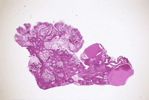
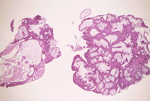
Gross pathology of the case:
A questionable mass and several areas of bullous edema near the dome of the urinary bladder were found by cystoscopy. The suspicious areas were biopsied and yielded the material being shown here.
Histopathology of the case:
|
|
|
|
|
|
|
|
| A. | B. | C. | D. | E. | F. | G. |
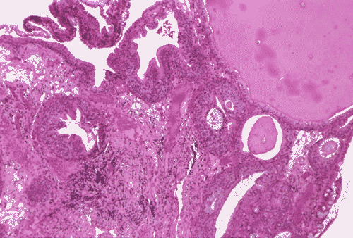
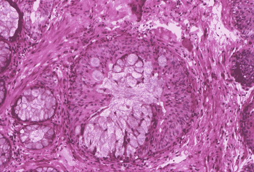
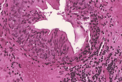
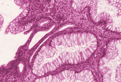
The
biopsy material showed polypoid fragments with cystically dilated glands lined
by columnar to cuboidal cells (Panel
A,
B, and
C).
Some of the glands were lined by transitional epithelium (Panel
D),
some were lined by a mixed transitional epithelium and mucin producing columnar
epithelium (Panel
E), and some
were lined entirely by mucin producing columnar epitheliuim (Panel
F)
that closely resembled colonic epithelium. A mild to moderate degree of
chronic inflammatory cell infiltration was also present. A mixed population of
mucin producing columnar epithelium with transitional epithelium was also noted
in many of the non-glandular surface epithelium (Panel
G).
No dysplasia was found.
| DIAGNOSIS: Polypoid
cystitis cystica et glandularis, intestinal type. |
Discussion: General Information Pathology Differential diagnosis
General Information
Cystitis
glandularis and cystica usually occurs in chronically irritated bladders such as
the non-functional bladder in paraplegics or in patients with long-term
catherization, as in our case, or stone.
1,
2
Von
Brunn’s nest refers to nests of transitional cells within the lamina propria
of the urinary bladder that arise from invagination of the overlying urothelium.
It is the most common reactive proliferative change within the urothelium
and is common enough to be considered a normal feature of the bladder mucosa by
some investigators. Cystitis glandularis is a common change that occurs most
often in the trigone. It is an inflammatory process that arises within and merges
imperceptibility with von Brunn’s nests. With cystic dilatation of the glands,
a progressive flattening of the lining cells occurs, and the lesion takes on the
form of cystitis cystica.
3,
4
Cystitis glandularis is microscopic or occurs as small mucosal nodules in most
occasions but it may occur as polypoid mass that suggests a neoplastic process
macroscopically and endoscopically.
The
typical type of cystitis glandularis contains simple cuboidal to columnar cells.
Small amount of mucin may be present but the amount is usually not impressive.
The intestinal type contains epithelium with intestinal metaplasia. These type
has numerous goblet cells and tubular epithelium that morphologically resemble
colonic epithelium and contains colonic type mucin
5,
6
.
Paneth cells metaplasia can also be seen in this type. Cystitis glandularis is
associated with increased risk of adenocarcinoma
1,
3,
4,
5,
7,
8.
Patients with extensive and diffuse intestinal metaplasia are particularly prone
to develop adenocarcinoma
8.
Differential diagnosis
The histopathologic diagnosis of cystitis glandularis and cystica rarely poses a real diagnostic problem. The most important distinction is between this benign lesion and adenocarcinoma. Cystitis glandularis and cystica usually lacks the nuclear atypia and stromal reaction seen in invasive adenocarcinoma. Furthermore, glands in cystitis glandularis are smooth in contour while those of adenocarcinoma are more irregular and give an infiltrative architectural pattern. Deep location, irregular contour, desmoplastic reaction, and nuclear atypia are features that suggest adenocarcinoma 9. Substantial mucin extravasation into the stroma can be seen in some cases of the intestinal type of cystitis glandularis, the distinction between cystitis glandularis and adenocarcinoma may be difficult in these cases 10, 11, 12.
Nephrogenic adenoma is another lesion with a higher incidence in previously manipulated or traumatized bladders and most of them are seen in adults. This condition is often associated with chronic cystitis. The inflammatory component may obscure the morphology. Histologically, they occur with tubular, cystic, polypoid, papillary, and diffuse pattern. The lining epithelium is cuboidal to low columnar with scant cytoplasm. The cells resemble renal tubules. In contrast to cystitis cystica, they are not surrounded by transitional epithelium. In nephrogenic adenomas, flat epithelium may be found in larger cysts ; cells with abundant clear cytoplasm and hobnail cells can also be found. Occasionally, nephrogenic adenoma may be associated with cystitis glandularis.
Reference:
Delnay
KM, Stonehill WH, Goldman H, Jukkola AF, Dmochowski RR.
Bladder histological changes associated
with chronic indwelling urinary catheter. J
Urol. 1999 161:1106-8; discussion 1108-9.
Fein
RL, Winton L, Gomez RR, Needell MH. Bladder calculi enveloped by
extensive cystitis glandularis: case report. J Urol. 1983
130:558-9.
Young,
RH. Pathology of the Urinary Bladder.
Churchill Livingstone, New York 1989.
Bostwick DG and Eble JN. Non-Neoplastic disorders of the urinary bladder in Urologic Surgical Pathology. Mosby, St. Louis, 1997.
Newbould
M, McWilliam LJ.
A
study of vesical adenocarcinoma, intestinal metaplasia and related lesions
using mucin histochemistry. Histopathology.
1990 17:225-30.
Wells
M, Anderson K. Mucin histochemistry of cystitis glandularis and
primary adenocarcinoma of the urinary bladder. Arch Pathol Lab Med.
1985 109:59-61.
Edwards
PD, Hurm RA, Jaeschke WH. Conversion of cystitis glandularis to
adenocarcinoma.
J Urol.
1972 108(4):568-70.
Lin
JI, Yong HS, Tseng CH, Marsidi PS, Choy C, Pilloff B. Diffuse
cystitis glandularis. Associated with adenocarcinomatous change. Urology. 1980 15:411-5.
Jacobs
LB, Brooks JD, Epstein JI. Differentiation of colonic metaplasia
from adenocarcinoma of urinary bladder. Hum Pathol. 1997
28:1152-7.
Eble
JN, Young RH. Carcinoma of the urinary bladder: a review of its
diverse morphology.
Semin Diagn Pathol.
1997 14:98-108.
Young
RH.. Pseudoneoplastic lesions of the urinary bladder and urethra:
a selective review with emphasis on recent information. Semin
Diagn Pathol. 1997
14:133-46.
Young
RH, Bostwick DG. Florid cystitis glandularis of intestinal type
with mucin extravasation: a mimic of adenocarcinoma. Am J Surg Pathol. 1996
20:1462-8.