| A 35 year-old Asian Man with
Multiple Small Nodules in the Neck. November, 2003, Case 311-2. Home Page |
O. Hans Iwenofu, M.D., Kar-Ming Fung, M.D., Ph.D. Last update: December 30, 2003.
Department of Pathology, University of Oklahoma Health Sciences Center, Oklahoma City, Oklahoma
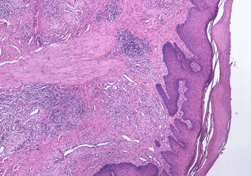
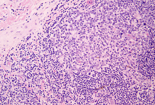
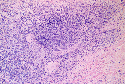
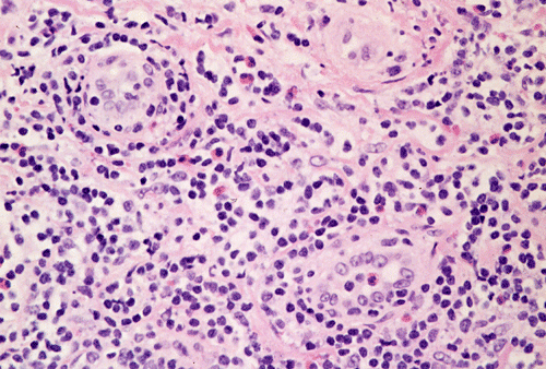
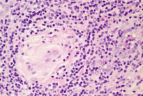
Clinical information: The patient was a 35 year old man who presented with multiple non-ulcerated small nodules in the preauricular area and the neck. As per the patient, the lesion had been there for an unknown but long duration. On physical examination, the cervical nodes are slightly enlarged. A biopsy of the skin lesion was taken. Representative areas are shown below:
|
|
|
|
|
||
| A. | B. | C. | D. |
Pathology of the case:
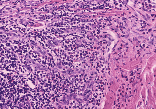
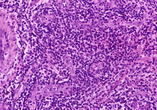
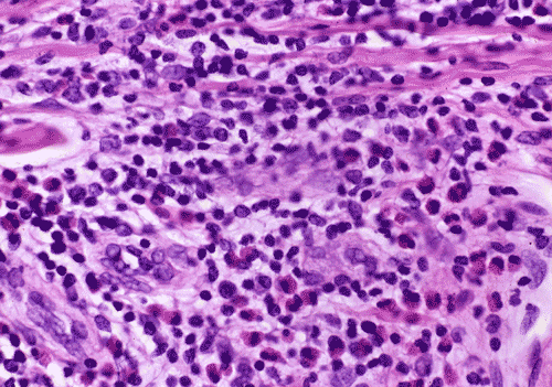
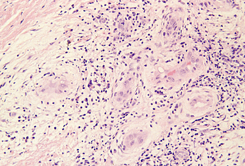
On low-magnification (Panel A), the subcutis is infiltrated by chronic inflammatory cells in the pattern of perivascular aggregates. On medium-magnification (Panel B), the infiltrate is composed predominantly of small lymphocytes without atypia and occasional eosinopils (arrow in Panel B; Panel C). There is a good number of blood vessels in this photo but there is no plump, epithelioid endothelial cells. The cytologic details are better demonstrated in high-magnification photos. Small clusters of eosinophils are present (Panel D).
| DIAGNOSIS: Kimura's disease. |
Discussion: General Information Pathology Pathogenesis Differential diagnosis Treatment
General Information
Kimura’s disease is a chronic inflammatory disorder of uncertain etiology. It
was first described in China in 1937 by Kim and Szeto as eosinophilic
lymphogranuloma 1. This entity became more
widely recognized as Kimura’s disease in 1948 after a systematic description
by Kimura et al who aptly described the condition as “an unusual granulation
combined with hyperplastic changes of lymphoid tissue"
2.
Kimura’s disease is most commonly seen in patients between 20-40 years of age
with a striking male predominance. Occurrence in children and elderly patients
can occur. Typically they occur as multiple cutaneous nodules in the head and
neck region particularly the preauricular regions. The patient often reports a
long duration of disease up to 10 years. Lymphadenopathy is present in over half
of the cases. Kimura’s disease is endemic among the oriental population with
few isolated reports in other parts of the world. Young and middle-aged Asian
males of Chinese and Japanese ancestry are primarily affected. Occasionally, it
is seen in other parts of the world
3.
It
is characterized by a triad of insidious onset of painless subcutaneous nodules
in the head and neck region, blood and tissue eosinophilia and markedly elevated
serum immunoglobulin levels
4.
It is an important cause of lymphadenopathy in Oriental patients. In the head
and neck areas, Kimura’s disease has been described in lymph nodes of the head
and neck region
4,
5,
parotid and submandibular gland
6,
oral mucosa
7,
auricle
8,
scalp
9
and orbit
10.
Outside the head and neck region, Kimura’s disease has been described in
axillary and inguinal lymph nodes
4,
spermatic
cord
11,
peripheral nerve
12
and other sites. Occasional cases are associated with renal diseases and
nephritic syndrome
13,
14,
15,
bronchial asthma
16,
17
and ulcerative colitis
18.
The histomorphology of Kimura’s disease is in cutaneous nodules is
characterized by intense infiltration of lymphocytes and plasma cells with a
variable number of lymphoid follicles with germinal centers. Typically, there is
a moderate to intense eosinophilic infiltration in the background with focal
eosinophilic abscess in some cases.
The involved lymph nodes shows marked hyperplasia of germinal centers that are
often well vascularized and contain Warthin-Finkeldey type polykaryocytes,
hyalinized vessels in the paracortex, variable amount of sinusal and
paracortical sclerosis, and deposition of proteinaceous material. There is also
increase in the number of plasma cells and mast cells in the paracortex.
Immuohistochemical stains would typically show Ig E reticular network in the
germinal centers
19.
The exact cause of Kimura’s disease remains an enigma. It has been speculated
that a viral or a parasitic trigger may cause a T-cell immunodysregulation or
induce an IgE-mediated type-1 hypersensitivity resulting in the release of
eosinophiliotrophic cytokines
20,
21,
15
but no incontrovertible evidence exists on these claims. Its occasional
association with with nephritic syndrome
22,
15,
bronchial asthma
23,
17
and ulcerative colitis
18
and deposition of IgE in lesional tissue
19
certainly would suggest an immunologic pathogenesis. Immunohistochemical
studies performed on skin, lymph nodes, and peripheral blood in kimura’s
disease have shown marked proliferation of human leucocyte antigen-DR CD4 cells
24.
Activated
CD4 cells of the TH2 phenotype can release cytokines such as granulocyte
macrophage colony-stimulating factor, tumor necrosis factor alpha,
interleukin(IL)-4 and IL-5 which would lead to high serum levels of IgE and
consequently marked eosinophilia
16.
There has been ultrastructural studies indicating the possibility that the slow
release of mediators or cytokines from mast cells by piecemeal degranulation may
contribute to the pathomechanism of Kimura’s disease
25.
Recently
there is an isolated report alluding to the presence of clonal T-cell population
in kimura’s disease
26.
Differential diagnosis
Until recently the distinction between atypical lymphoid hyperplasia with
eosinophilia (ALHE) and Kimura’s disease has been quite confusion. This
two diseases are now recognized as two separate entities in the pioneering work
by Rosai
27.
Other
observations also alluded to the fact that inspite of their common similarities
they still remain clinical and morphological different
5,
28,
29,
30.
 Click thumbnails
to see pictures.
Click thumbnails
to see pictures.
The treatment of Kimura’s disease remains a challenge
32,
33
Following
initial presentation, surgical excision with diagnostic intent might be curative
but recurrence is common
20.
Other treatment options include radiation, systemic corticosteroids,
cyclosporine and pentoxyfylline have been tried with variable results
32,
33,
34.
Radiation is typically reserved for cases refractory to medical treatment or in
large tumors where surgery is not technically feasible
34.
Reference:
Kim
HT, Szeto C. Eosinophilic hyperplastic lymphogranuloma, comparison with
Mikulicz’s disease. Chin med J. 1937 23:699-700.
Kimura T, Yoshimura S, Ishikaura E. Unusual granulation combined with hyperplastic changes of lymphatic tissue. Trans Soc Pathol Jpn. 1948;37:179-180.
Pamaraju N, Khalifa SA, Darwish A, Paulose KO, Ahmed N, Yousif H. Kimura’s disease. J Laryngol Otol 1996;110:1084-1087.
Kuo TT, Shih LY, Chan HL. Kimura’s disease. Involvement of regional lymph nodes and distinction from angiolymphoid hyperplasia with eosinophilia. Am J Surg Pathol. 1988;12:843-854.
Tham KT, Leung PC, Saw D, Gwi E. Kimura’s disease with salivary gland involvement. Br J Surg.1981;68:495-497.
Hongcharu W, Baldassano M, Taylor CR. Kimura’s disease with oral ulcers: response to pentoxyfylline. J Am Acad Dermato. 2000;43:905-907.
Chan KM, Mok JS, Ng SK, Abdullah V. Abdullah V. Kimura’s disease of the auricle. Otolaryngol Head Neck Surg. 2001;124:598-599.
Jambhekar NA, Borges AM, Saxena R, Parikh DM, Soman CS. Angiolymphoid hyperplasis with eosinophilia (Kimura’s disease). Report of a large sized lesion. J Surg Oncol 1991;47:206-208
Buggage RR, Spraul CW, Wojno TH, Grossniklaus HE. Kimura disease of the orbit and ocular adnexae. Surv Opthalmol.1999;44:79-91.
van Gulik TM, Jansen JW, Taat CW. Kimura's disease in the spermatic cord, an unusual site of a rare tumor. Neth J Surg. 1986 Jun; 38(3): 93-5.
Lee YS, Ang HK, Ooi LL, Wong CY. Kimura's disease involving the median nerve: a case report. Ann Acad Med Singapore. 1995 May; 24(3): 462-4.
Chartapisak W, Opastirakul S. Steroid-resistant nephrotic syndrome associated with Kimura's disease. Am J Nephrol. 2002 Jul-Aug; 22(4): 381-4.
Romao JE, Saldanha LB, Ianez LE, Sabbaga E. Recurrence of focal segmental glomerulosclerosis associated with Kimura's disease after kidney transplantation. Am J Kidney Dis. 1998 Mar; 31(3): E3.
Rajpoot DK, Pahl M, Clark J. Nephrotic sundrome associated with Kimura disease. Pediatr Nephrol.2000;14:486-48.
Tsukadaira A, Kitano K, Okubo Y, Horie S, Ito M, Momose T, Takashi S, Itoh S, Kiyosawa K, Sekiguchi M. A case of pathophysiological study in Kimura’s disease: measurement of cytokines and surface analysis of eosinophils. Ann Allergy Asthma Immunol.1998;81:423-427.
Okudaira H, Hongo O, Ogita T, Haida M, Yamauchi N, Miyamoto T. Serum IgE and IgE antibody levels in patients with bronchial asthma, atopic dermatitis, eosinophilic granulomas of the soft tissue (Kimura's disease) and other diseases. Ann Allergy. 1983 50: 51-4.
Sugaya M, Suzuki T, Asahina A, Nakamura K, Ohtsuki M, Tamaki K. Kimura's disease associated with ulcerative colitis: detection of IL-5 mRNA expression of peripheral blood mononuclear cells and colon lesion. Acta Derm Venereol. 1998 Sep; 78(5): 375-7.
Motoi M, Wahid S, Horie Y, Akagi T. Kimura’s disease: clinical, histological, and immunohistochemical studies. Acta Med Okayama.1992;46:449-455.
Armstrong
WB, Allison G, Pena F, Kim JK.
Kimura’s disease: two case reports and a literature review. Ann Otol
Rhinol Laryngol. 1998;107:1066-1071.
Chusid MJ, Rock AL, Sty JR, Oechler HW, Beste DJ. Kimura’s disease: an unusual cause of a cervical tumor. Arch Dis Child. 1997;77:153-154.
Chartapisak W, Opastirakul S. Steroid-resistant nephrotic syndrome associated with Kimura's disease. Am J Nephrol. 2002 22 381-4.
Tsukagoshi H, Nagashima M, Horie T, Oyama T, Yoshii A, Sato T, Iizuka K, Dobashi K, Mori M. Kimura's disease associated with bronchial asthma presenting eosinophilia and hyperimmunoglobulinemia E which were attenuated by suplatast tosilate (IPD-1151T). Intern Med. 199837: 1064-7.
Tabata H, Ishikawa O, Ohnishi K, Ishikawa H. Kimura’s disease with marked proliferation of HLA-DR+ CD4+ T cells in the skin, lymph node, and peripheral blood. Dermatology.1992;184;145-148.
Aoki M, Kawana S. The ultrastructural patterns of mast cell degranulation in Kimura’s disease. Dermatology.1999; 199(1):35-9.
Chim CS, Fung A, Shek TW, Liang R, Ho WK, Kwong YL. Analysis of clonality in Kimura’s disease. Am J Surg Pathol.2002 Aug;26(8):1083-6.
Rosai J. Angiolymphoid hyperplasia with eosinophilia of the skin: Its nosological position in the spectrum of histiocytoid hemangioma. Am J Dermatopathol 1982 4:175-178.
Chan JK, Hui PK, Ng CS, Yuen NW, Kung IT, Gwi E. Epitheliod heamngioma(angiolymphoid hyperplasia with eosinophilia) and Kimura’s disease in Chinese. Histopathology 1989 15:557-574.
Chun SI, Ji HG. Kimura’s disease and angiolymphoid hyperplasia with eosinophilia: clinical and histopathological differences. J Am Acad Dermatol.1992;27:954-958.
Seregard S. Angiolymphoid hyperplasia with eosinophilia should not be confused with Kimura’s disease. Acta Opthlmol. Scand 2001;79:91-93.
Fetsch
JF, Weiss SW.
Observations concerning the pathogenesis of epitheloid
hemangiomas(angiolymphoid hyperplasia). Mod Pathol 1991 4:449-455.
Olsen TG, Helwig EB. Angiolymphoid hyperplasia with eosinophilia. A clinicopathologic study of 116 patients. J Am Acad Dermatol.1985;12:781-796.
Kim GE, Kim WC, Yang WI, Kim SK, Oh WY, Suh HS, Hahn JS, Park CS. Radiation treatment in patients with recurrent Kimura’s disease. Int J Radiat Oncol Biol Phys.1997;38:607-612.