
| A 23 year-old Man
with a Mandibular Mass. August, 2004, Case 408-1. Home Page |
Glen D. Houston, D.D.S.1, Kalliopi Petropoulou, M.D. 2, Kar-Ming Fung, M.D., Ph.D. 1 Last Update September 30, 2004.
Department of Pathology1 and Department of Radiology 2, University of Oklahoma Health Science Center, Oklahoma City, Oklahoma.
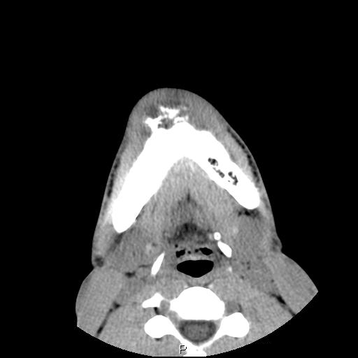
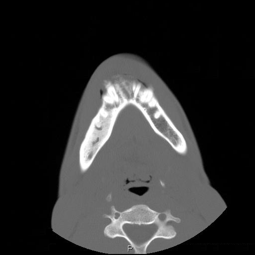
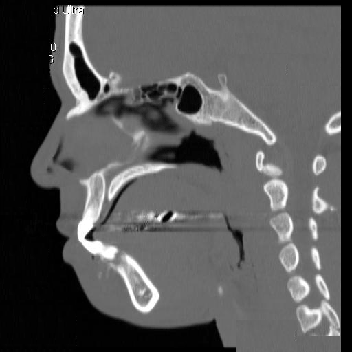
Clinical information: The patient was a 23 year-old man who went to see the dentist because of dull pain, swelling of the chin, and loosened lower front teeth. He was referred to this hospital after initial examination. A CT scan was performed and and a biopsy was taken. Representative images are presented here. Panel D is a sagittal reconstruction from axial CT scans. A resection was subsequently performed.
|
|
|
|
|
||
| A. | B. | C. | D. | ||
|
|
|
|
|||
| G. | H. | I. |
CT scan: On the CT scan images (Panel A, B, C, and D), two lesions are present. There is a lesion at the mental protuberance that appears to have penetrated the cortex and invaded into the soft tissue. The patchy hyperdensity strongly suggests that this lesion has mineralize elements ( †in Panel B). The border of the lesions in the mandible is poorly defined. The penetration of the bone and extension into the soft tissue is best appreciated in the sagittal reconstruction in (Panel D). These features are highly suggestive of a malignant neoplasm that originates from the bone with extension into surrounding soft tissue. The hyperdensity of the mass suggests bone formation and the radiologic features are suggestive of an osteosarcoma. In the right body of the mandible, there is a diffusely expansile lesion with some hazy hyerdensity (Ú in Panel B). The cortical bone overlying this lesion is intact. This lesion may represent extension of the osteosarcoma into the body. However, it may also represent a co-existing fibrous dysplasia.
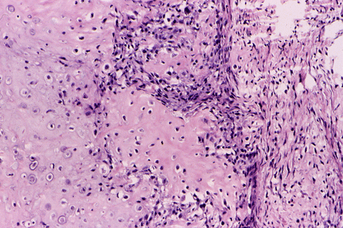
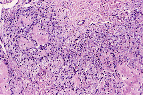
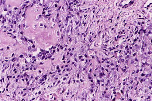
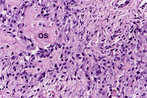
Pathology of the case: An incisional biopsy yielded several small fragments of gritty, tan to translucent tissue. The histopathology varies in different parts of the tumor. In some areas, the lesion is composed predominantly of islands of neoplastic cartilage with focal osteoid formation and rimmed by spindle cells (Panel E). In other areas, densely packed spindle cells proliferation admixed with hypocellular fibrous tissues is present (Panel F). On high magnification, the spindle cells appear to be pleomorphic and hyperchromatic (Panel G). Mitotic figures are present but not numerous. No atypical mitoses are noted. Small foci of osteoid are best appreciated at higher magnification ("OS" in Panel G). It appears that this tumor has arisen from the medullary cavity and is best classified as a central osteosarcoma.
| DIAGNOSIS: Osteosarcoma, chondroblastic type. |
Discussion: General Information Radiology Pathology Differential diagnosis
General Information
Osteosarcomas of the jaws comprise about 10% of all the osteosarcomas. They are different from osteosarcomas of the long bones in several aspects. Although they can occur from children to elderly, they are seen most commonly in the 3rd and 4th decades which is 10 to 15 years older than the mean age for osteosarcomas of the long bones. In contrast to osteosarcoma of other locations, the incidence of osteosarcoma of the jaws does not have a biomodal distribution. They are somewhat more common in males. The maxilla and mandible are involved in comparable frequency. The alveolar ridge of the maxilla and the body of the mandible are the most common site, they are quite uncommon in other craniofacial regions. Osteosarcomas of the skull are related to Paget's disease. Interestingly, osteosarcomas almost always arise from craniofacial bones with intramembranous ossification and practically never occur in the bones of the base of the skull that are formed by endochondrial ossification. In contrast, chondrosarcomas can arise from the bones of the skull base.
While pain is the major complain in osteosarcomas arising in other parts of the body, swelling is the major complain in osteosarcoma of the jaws. Other symptoms include paresthesia and loosening of teeth. Nasal obstruction, symptoms mimicking sinusitis, and facial swelling can occur when the osteosarcoma arises in the maxilla. The duration of symptoms is partly related to the rate of growth of the tumor. A small number of osteosarcomas of the jaws grow rather slowly and the initial symptoms may mimic teeth problems. The duration of symptoms in these patients may also be long.
The level of serum alkaline phosphatase is frequently elevated in patients with osteosarcomas. The level is particularly high in patients with large tumor burden and osteosarcomas with prominent osteoblastic differentiation. Elevation of alkaline phosphatase level after surgery indicates metastatic or persistent disease. Serum level of alkaline phosphatase is also one of the effective means to detect recurrence.
In general, osteosarcomas of the jaws have a better prognosis than those arising from the long bones partly because low-grade osteosarcomas are more common in the jaws. High grade osteosarcomas of the jaws also behave aggressively. The tumor can occur as central (intramedullary) osteosarcoma, periosteal osteosarcoma, and parosteal osteosarcoma. Central osteosarcomas have the worst prognosis. Juxtacortical (parosteal and periosteal) osteosarcomas have a better prognosis but these tumors are rare. Osteosarcomas of the craniofacial regions have a high local recurrence rate of 50% and this is the most important reason for therapeutic failure. Metastases are less frequent than their long bone counterparts and they occur predominantly as lung metastases within the first two years after surgery.
Radiologically, the amount of calcified content determined the overall density of the lesion. It can vary from dense sclerosis, to a mixed sclerotic and radiolucent lesion as illustrated in this case, to entirely radiolucent. The destructive nature of the tumor leads to a gradual transition of tumor to normal bone, which makes the border of the tumor ill-defined. As a rule, the intramedullary extension is typically more extensive than is estimated on plain radiographs. The classic "sunburst" changes of the periosteum is present in about one-fourth of the cases. The tumor can destroy the cortex and penetrate into the soft tissue as in this case. Root resorption often lead to tapered narrowing of the root and is described as "spiking" resorption. Tumor infiltration along the periodontal ligament space leads to a symmetric widening of the periodontal ligament space around a tooth or several teeth on bite-wing radiograph. This is an important early radiological sign for osteosarcoma. Such widening, however, is not specific for osteosarcoma and is also seen in other malignancies infiltrating into the periodontal ligment space.
Macroscopically, osteosarcoma appears as an invasive, expansile mass. Most of them arise from the medullary cavity (central osteosarcoma). A small number of them arise in the juxtacortical location (parosteal and periosteal osteosarcoma). The color of the cut surface is very heterogeneous. The consistency may vary from highly calcified, hard area to soft, friable, and hemorrhagic.
Osteosarcoma is a malignant tumor characterized by direct formation of bone or osteoid by the proliferating neoplastic cell. Other than sharing this characteristic, osteosarcoma is one of the most heterogeneous tumor from the histopathologic perspective. The histologic features vary from different areas of the same tumor and among different tumors. In general, several major patterns are recognized: osteoblastic, chondroblastic, fibroblastic, giant-cell rich, small cell, telangiectatic, and others. The amount of osteoid and ossified tissue can vary tremendously and it is not uncommon that it can only be found after careful search. The osteoid can form well-defined structures that mimic trabecular bone or as irregular deposits. The current trend is to classify osteosarcomas into low- and high-grade tumors. It is the histologic grade rather than tissue pattern that has greatest bearing on prognosis.
Similar to osteosarcomas arising from the flat bones and ribs (e.g., osteosarcomas of the pelvic bones), about 30% of the osteosarcomas of the jaws are chondroblastic type and contain a substantial amount of cartilage. Lobules of neoplastic cartilage may dominate the histologic picture in some cases and osteoid production by the neoplastic cells may occur only focally. The cartilaginous differentiation can be heterogeneous and represent all stages of cartilaginous matrix formation. Stigmata of malignancies including nuclear pleomorphism, mitosis, atypical mitotic figures, and necrosis can all be seen in osteosarcomas.
Differential diagnosis
Several lesions closely mimic osteosarcoma of the jaws. The differential diagnoses include fibromyxoid chondroma, fibro-osseous lesions, chondrosarcoma, aneurysmal bone cyst, and central giant cell granuloma.
Fibromyxoid chondroma is a benign tumor characterized by lobules of myxoid and chondroid material separated by zones of cellular spindle-shaped or rounded cells. About 75% of these tumors occur in the 1st and 2nd decades of life and they are most commonly seen in long bones. Typically, these lesions occur as eccentrically located radiolucent lesions in the metaphysis with a scalloped and sclerotic inner wall. The epiphyseal line is only rarely invaded. Macroscopically, they have a bluish, translucent cut surface. Histologically, lobulated areas of spindle-shaped or stellate cells with abundant myxoid or sometimes chondroid material intermingled with areas of more densely packed spindle-shaped or rounded cells and a variable number of multinucleated giant cells may be observed. Mitotic figures were generally lacking. The cells in the chondroid area usually maintain a spindle morphology instead of the round nuclei found in chondrocytes. Occasional pleomorphic cells can be present. .
Fibro-osseous lesions contain both a fibrous and osseous component. This is a collective term for fibrous dysplasia, ossifying fibroma, and cementifying fibroma. These lesions are radiologically different from that of osteosarcoma. Although both fibrous and osseous components are present, a lack of the stigmata of malignancy separates them from osteosarcoma.
Separation of chondrosarcoma from chondroblastic type of osteosarcoma of the jaw can be a challenge. The key features is to identify the osteoid forming component. In fact, whether cartilage dominant osteosarcoma of the jaw should be classified as osteosarcoma or chondrosarcoma remains a good academic question.
Central giant cell "reparative" granuloma is a benign non-neoplastic giant cell lesion arising centrally within the jaws. The name is a misnomer since these lesions do not contain granulomas and are not necessary reparative. They are locally destructive and usually affect children and young adults, predominantly females. They are most frequently seen in tooth bearing areas of the mandible, followed by the maxilla, and infrequently seen in small bones of the fingers. Radiographically, they present as osteolytic lesions similar to aneurysmal bone cysts. Histologically, there are lobules of connective tissue composed of fibrobrast-like spindle cells, collagen, and foci of hemorrhage within which multinucleated giant cells of osteoclast type are clustered. Hemosiderin deposits and reactive woven bone may be present. These lesions are histologically indistinguishable from brown tumors associated with hyperthyroidism. In general, these lesions lack the stigmata of a sarcoma. It should also be noted that biopsy material that are obtained from the periphery of an osteosarcoma my mimic central giant cell granuloma.
Aneurysmal bone cyst can be distinguished from osteosarcoma in most occasions both radiographically and pathologically. It should be noted that a small number of osteosarcomas are associated with aneurysmal bone cysts. A careful examination of the surgical material is mandatory.
Further Reading: