PAS
PAS
| A 41 year-old Woman with a
History of Leukemia and now Presented with Chronic Sinusitis. October, 2008, Case 810-1. Home Page |
Douglas W. Warden, M.D.1, Jose A. Sanclement, M.D.2, Gregory L. Blakey, M.D.1, Kar-Ming Fung, M.D. Ph.D.1
1 Department of Pathology, and 2 Department of Otorhinolaryngology, University of Oklahoma Health Science Center, Oklahoma City, OK. Last update: October 31st, 2008.
Clinical information: The patient was a 42 year-old woman with a history of acute promyelocytic leukemia status post Atra and induction chemotherapy with Idarubicin and ARA-C was admitted to the hospital for consolidation therapy. During her hospitalization she developed mucositis which gradually progressed for a few weeks. Her symptoms included sinus pain (left significantly greater than right), sinus drainage, mouth pain (particularly on the soft palate), and facial swelling. Computerized tomography (CT) scan revealed destructive changes of the hard palate and maxilla, and surgical intervention was required. Intra-operative findings included bilateral diffuse necrosis of the nasal cavities, with significant destruction of the nasal septum, left nasal turbinate, and bony nasal floor. Specimens were sent for frozen section, permanent section, and fungal cultures. The patient received antifungal therapy and required repeated sino-nasal debridement. There has been no further extension of fungal infection. Following are images from permanent section.
|
|
|
|
|
|
|
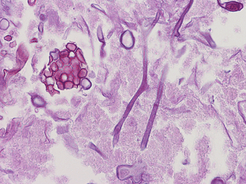
| A. | B. | C. |
D. PAS |
E. PAS |
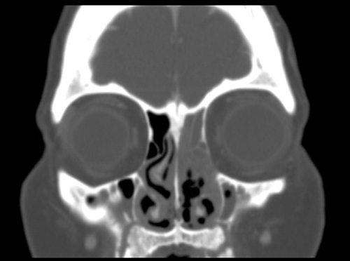
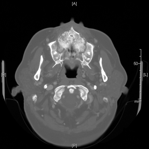
Image of the Case: CT scan, coronal view, of the sinuses reveals an opacified left ostiomeatal unit on the left and thickening of the mucosa on the right (Panel A). CT scan, axial view. The hard palate and maxilla show destructive changes and fractures causing a “moth-eaten” appearance (Panel B).
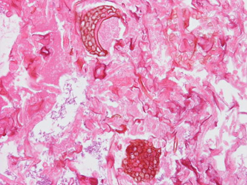
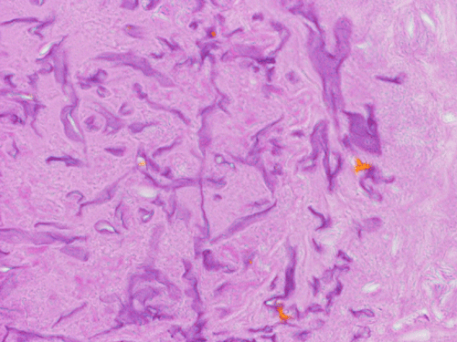
Pathology of the Case: Multiple tissue samples were from several locations of the sinuses were rerceived. The specimen was in the form of small aggregates of irregularly shaped, tan brown, necrotic debri with clotted blood and purulent material. Admixed with the soft tissue were small fragments of bone. Frozen section of “left nasal contents” reveals fungal organisms with hyphae and nonviable collagenous tissue, suspicious for invasive fungal infection. On permanent sections, about 99% of the tissue is composed of necrotic debri. There is an abundance of fungal organisms. While some of the organisms are scattered, there are areas suggestive of an angioinvasive pattern. These organisms may well be recognized by hematoxylin and eosin stain but also well demonstrated by PAS and GMS stain. The organisms contain pauciseptate, “ribbon-like,” irregular hyphae with acute and right-angle branching. Associated with the hyphae are globus-shaped, pigmented sporangia and columella (Panel C, D, and E). The hyphae are wide and here show acute angle branching. Occasional septum can be seen (Panel D and E). The sporangium on the left shows angulated sporangiospores (Panel C and E). These features are characteristic of the class Zygomyycetes, order Mucorales. This order includes the Rhizopus and Mucor species. Fungal culture identified a Rhizopus specie as well as Aspergillus. The pathologic features are most compatible with a sino-nasal mixed fungal infection in an immunocompromised host.
| DIAGNOSIS: Invasive fungal infection with Invasive mucormycosis, Rhizopus species, and Aspergillus. |
Discussion:
Mucormycosis (also known as zygomycosis) is caused by a group of fungi including Rhizopus, Rhizomucor, Cunninghamella, Apophysomyces, Saksenaea, Absidia, Mucor, Syncephalastrum, Cokeromyces, and Mortierella. Among these, Rhizopus is the most common cause. These organisms are ubiquitous, present in soil, compost, decaying vegetable matter, and water. Typically, infection is associated with diabetic patients (particulary during diabetic ketoacidosis), but immunocompromised patients are also at significant risk. Most often, infection involves the paranasal sinuses and from there may rapidly invade the orbit and brain. The organisms characteristically invade the blood vessels where they can cause thrombosis, hemorrhage and eventually infarction.
All Zygomycota share similar morphologic features with broad, thin-walled, pauciseptate hyphae. Typically the different genera can be separated by the appearance of their sporangia and sporangiophores grown in culture media. In this case, the globus shape and brown pigmentation of the sporangia along with the angulated shape and striations of the sporangiospores are consistent with the diagnosis of Rhizopus species. In the rare instance that these features are identified on tissue section, a presumptive diagnosis can be made. This diagnosis should be followed up by fungal culture confirmation. Diagnosis and management of fungal infection in immunocompromised patients often requires a multidisciplinary approach. Careful histologic evaluation may provide early identification of mucormycosis and save valuable time in directing focused therapy.
References:
Note: This case has been presented as a poster at the 2008 College of American Pathologist (CAP) Annual Meeting.