

| A 36 year-old Woman with a
Swelling in her Left Neck. October, 2008, Case 810-2. Home Page |
Asish Bains, M.D. , Elizabeth Gillies, M.D. Last update: October 31, 2008.
Department of Pathology, University of Oklahoma Health Sciences Center, Oklahoma City, Oklahoma
Clinical information: The patient is a 36 year-old woman who presented with a 2 cm swelling in the left side of her neck. The mass was not tender, warm, or painful. The overlying skin was unremarkable. A CT scan was performed and showed a 1.8 x 1.2 cm relatively non-enhancing lesion within the carotid sheath displacing the carotid artery anterioriorly. The clinical and imaging features yielded an empirical diagnosis of an enlarged lymph node. The patient had no history of lymphoma, leukemia, or head and neck tumor. The mass was removed surgically in its entirety and was 2.5 cm in maximum dimension, well encapsulated, located at the level of the carotid bifurcation just lateral to the carotid artery and superiorly extending almost up to the level of the tonsils. The mass did not appear to be in continuity with the jugular vein or the carotid artery. It was not a part of the vagus nerve or sympathetic chain. The followings are representative images.
|
|
|
|
|
|
|
|
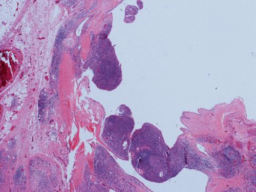
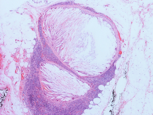
| A. | B. | C. | D. | E. | F. | Scanned slide |
Gross Pathology: The specimen is 4.0 x 1.2 x 1.2 cm, pink-tan to red, soft to firm and nodular mass. It appears to be cystic on cut section and has multiple lobules ranging from 0.2 to 0.7 cm in the maximum diameter. The cystic spaces contain sero-sanguinous fluid. The specimen is serially sectioned and entirely submitted for histologic examination.
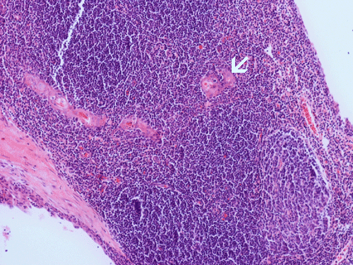
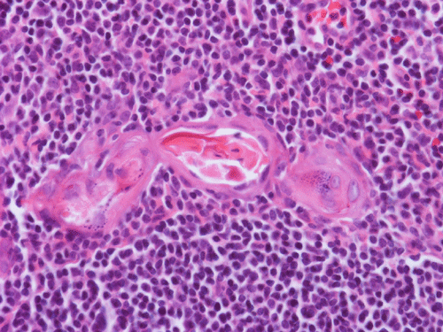
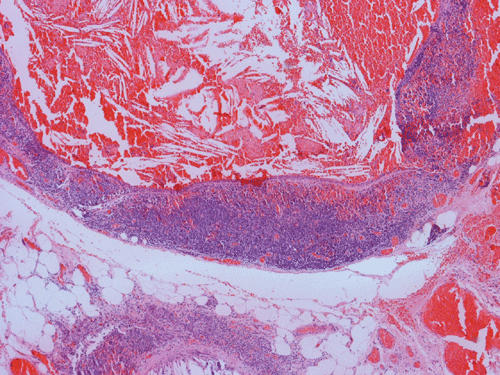
Histopathology: The lesion is encapulated cystic structure with the wall lined by nodules of lymphoid tissue (Panel A and B) that are devoid of any evidence of hematopoiecic malignancy. Within this lymphoid tissue are small pink islands (Arrow in Panel C) and on higher magnification (Panel D) these islands are squamous cell nests. These islands are Hassell corpuscles and the lymphoid tissue is in fact thymic tissue. The cyst appears to have sustained repeated hemorrages. Both recent hemorrhage (Panel E) and cholesterol clefts consistent with resolved hemorrhage (Panel F) are present.
| DIAGNOSIS: Cervical thymic cyst |
Discussion:
General Information:
Thymic cysts are rare benign causes of lateral neck masses. They are usually found in children and are asymptomatic in most cases. Approximately 150 cases 1 have been reported in the literature. Considering mostly benign nature of the lesion, they are underreported and probably occur more frequently. Clinically, most cases present as unilateral asymptomatic neck masses, commonly on the left side of the neck and 75% of the patients present before 20 years of age 2. Only 6 to 10% of the patients develop dysphagia, dyspnea, stridor, cervical pain, hoarseness and/or vocal cord paralysis 1, 3, 4.
Preoperatively, they are often mistaken as other processes such as lymphangiomas or branchial cleft cysts. The diagnosis relies on identification of Hassall’s corpuscles within a lymphoid background. Fine needle aspiration often yields a population of benign lymphocytes but not Hassall’s corpuscles. A correct preoperative diagnosis is not often achieved. In some cases the development of a mass may be noticed rather abruptly and, not uncommonly, resulting secondary to infection or hemorrhage in the cyst.
Approximately 50% have mediastinal extention 1, 5. In such cases where mediastinal extension is present, Valsalva maneuver will produce transient increase in size of the mass due to either vascular engorgement or increased intrathoracic pressure.
Pathogenesis and Embryology:
Many structures of the head and neck region arise from the pharyngeal pouches and pharyngeal arches. The thymus embryologically arises from the 3rd pharyngeal pouch or infrequently from the ventral portion of the fourth pharyngeal pouch. The third pharyngeal pouch gives rise to paired primordia of the thymus gland during the 6th week of fetal life. Following the fusion of the thymic primordia, the thymus becomes a mass of densely packed endodermal cells. By the 9th week of life the thymic tissue descends below the level of the clavicle. By the 10th week of life, this mass will be broken up by the invading mesenchyme into a nest of cells or a cytoreticulum. The thymic corpuscles of Hassall appear to be derived from outgrowth of endodermal epithelium. If the superior end of the thymic analage fails to regress in the eight week of embryonic life a sequestered solid or cystic nodule of tissue may be left along the course of migration and line of descend. The cervical thymic lesions can be categorized based on anatomic location and nature of tissue present into accessory cervical thymus, cervical thymic cyst, undescended cervical thymus, persistent thymic cord, cervical extension of mediastinal thymus, and ectopic thymus 2.
Speer 6 in 1983 postulated five possible theories on the pathogenesis of development of thymic cysts. Thus thymic cysts may represent: (1) embryonal remnants of the thymic tubules, thymopharyngeal ducts or branchial cysts, (2) degenerating Hassall’s corpuscles, (3) neoplastic processes in lymphoid, reticular or connective tissues, (4) sequestration products formed during the pathologic involution of the gland, and (5) lymph vessels, blood vessels or connective tissue in various stages of thymic development, hypo- or hyperplastic. These hypotheses do not exclude each other; on the contrary each one of these can be the basis for the different histological forms of cervical thymic cyst described. The most commonly accepted explanation currently is based of congenital or acquired theory; congenital being the persistence of the thymopharyngeal duct and the acquired being that thymic cysts develop from degeneration of Hassall’s corpuscles within remnants of ectopic thymic tissue.
Pathology:
Cervical thymic cysts can vary in size up to few centimeters, may be unilocular or multilocular. The lateral location is a good clue to support the diagnosis. The cyst contents vary from clear serous fluid to hemorrhagic or purulent fluid. They may contain semi-solid, gelatinous, necrotic debris, blood or cholesterol crystals (like this case). Thymic parenchyma, lymphoid tissue and Hassall’s corpuscles found within the cyst wall are considered pathognomic findings. The cyst wall may be devoid of epithelial lining or may have cuboidal, stratified or ciliated epithelium. Identification of the Hassall’s corpuscles is the key to correct diagnosis and sometimes they may not be abundant. Secondary changes due to hemorrhage are also common findings. Sometimes, pressure necrosis may lead to granulation tissue. No ectopic thyroid tissue should be present. The diagnosis is usually straight forward.
Reference:
Cigliano B, Baltogiannis N, De Marco M, Faviou E, Antoniou D, De Luca C ervical thymic cysts. Pediatr Surg Int 2007 23:1219-1225.
VK Shukul VK, Jha A, BG Shankar. Cervical thymic cyst NJAFI 2004; 60:204-205.
Burton EM, Mercado-Deane MG, Howell CG, Hatley R, Pfeifer EA, Pantazis CG, Chung C, Lorenzo RL. Cervical thymic cysts: CT appearance of two cases including a persistent thymopharyngeal duct cyst. Pediatr Radiol 1995 25: 363-365.
Terzakis G, Louverdis D, Vlachou S, Anastasopoulos G, Dokiankis G, Tsikou-Papafragou A. Ectopic thymic cyst in the neck. J Laryngol Otol 2000 114:38-320.
Hendrickson M, Azarow K, Ein S, Shandling B, Thorner P, Daneman A Congenital thymic cysts in children – mostly misdiagnosed. J Pediatr Surg 1998 33:821-825.
Speer FD. Thymic cyst: report of a thymus presenting cysts of three types. NY Med Coll Flower Hosp Bull 1938 1:142-150.