The first research effort that I initiated upon joining the OB/GYN Department was to understand the molecular and cellular changes that occur when a normal cell progresses to a preneoplastic tumor and on to a metastatic lesion. Cervical cancer was an appropriate model to start with because of the preneoplastic lesions induced by Human Papillomavirus (HPV) could be used for comparison with normal and cancerous cells. High risk HPV types are etiologic agents found in the vast majority of cervix tumors. They induce genetic instability leading to cancer. I utilized the skin equivalent model to develop a model of cervical tumor progression that compared the stages of normal, HPV-immortalized, low grade and high grade cervical tumors. Unlike conventional monolayer tissue culture, this organotypic model generates 3-dimentional tissue that can be evaluated with histology and immunohistochemistry. My article in Tissue and Cell 2: 269-274,1995 was the first demonstration that organotypic cultures of cancer cell lines could express proliferation and differentiation markers in patterns that represent the grade of tumor from which they were derived, thus validating the model system. Decreased expression of an oncogene (epidermal growth factoor receptor (EGF-R)) and a proliferation markeer (Ki-r7/myb) by retinoid treatment of these cervix cancer cultures is reported in Gynecologic Oncology 66: 114-121,1997 and J. Lower Genital Tract Disease, 3: 1-5, 1999, respectfully.
|
Organotypic cultures of HPV-immortalized preneoplastic keratinocytes called PPE-4 (A and C) and a cervical tumor cell line derived from a low grade tumor (CC-1) (B and D) were stained with an antibody to involucrin, a marker of differentiation (A and B), or with an antibody to Proliferating Cell Nuclear Antigen (PCNA), to identify actively proliferating cells (C and D). The PE-4 cultures exhibited characteristics typical of a preneoplastic lesion, in that proliferating cells are located in the basal and parabasal layers (C) and that involucrin is expressed focally (A). The CC-1 culture is typical of a low grade lesion, with proliferating cells located in all cell layers, but not in all cells (D) and involucrin is expressed focally (B). |
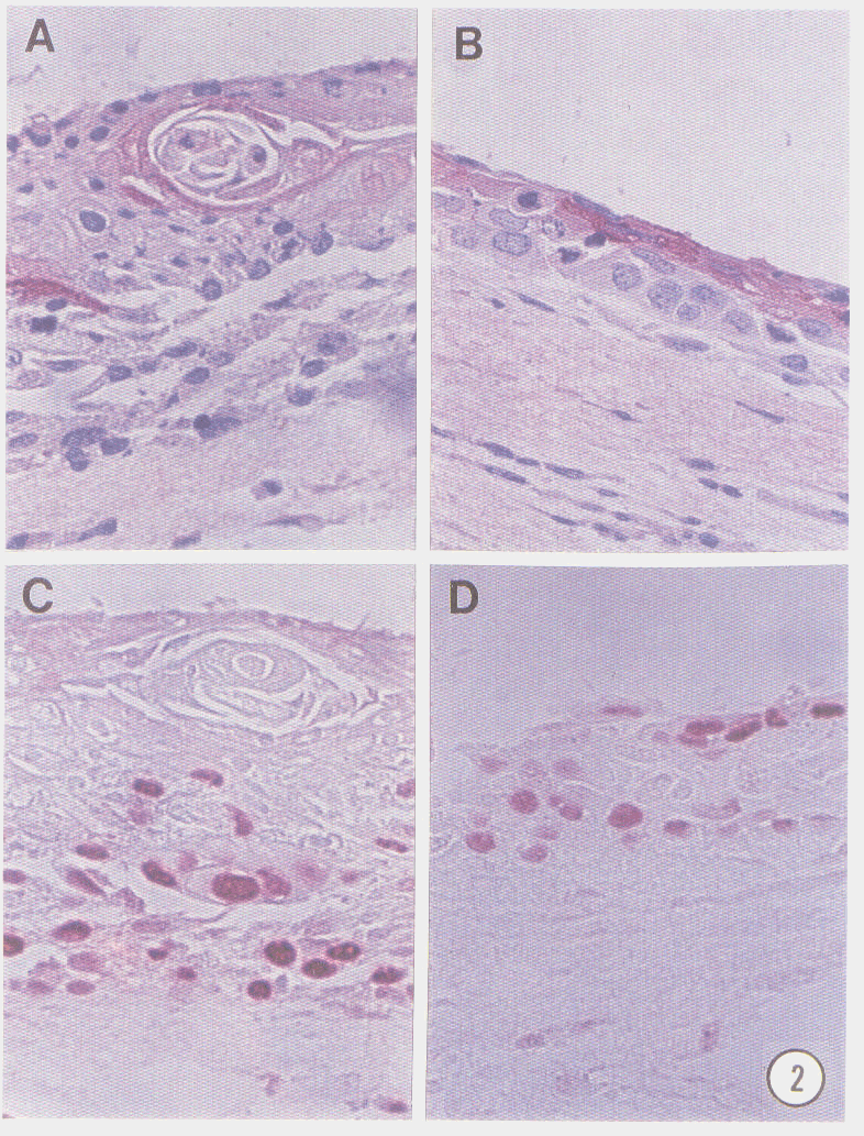
The SiHa and HeLa cervical tumor cell lines were also grown in organotypic culture and exhibited characteristics of high grade tumors. PCNA was expressed by all cells throughout the entire thickness of the culture and in patterns representative of all stages of the cell cycle. Neither cell line exhibited expression of differentiation which was evaluated by involucrin staining for squamous differentiation which was evaluated by involucrin staining for squamous differentiation and mucicarmin staining for adenocarcinoma differentiation.