BIOMECHANICAL EXAM FOR LE ORTHOSES - LAB

Each lab group needs:
Upon completion of this lab, students can:

Determining Subtalar Joint Neutral (Prone Examination)

The STJ is neutral when it is neither pronated or supinated. The neutral position is defined operationally in at least two ways.

Let ankle and foot hang over end of table. Place the calcaneus in the frontal plane by positioning the opposite hip in flexion, abduction and external rotation.

  1. Measuring Subtalar Joint Range of Motion

    With a skin pencil, draw a line that bisects the middle third of the calcaneus.

    Draw another line that bisects the distal third of the leg.

    Place one arm of a goniometer along either of these lines, keeping the goniometer's axis between the malleoli in the frontal plane.

    "Lock the forefoot" by grasping the fourth and fifh metatarsal heads and moving them dorsally. Maximally supinate the subtalar joint, realign the goniometer's calcaneal arm, and read the degrees of inversion.

    Maximally pronate, realign goniometer, and read degrees of eversion.

  2. Finding Subtalar Joint Neutral

    • Qualitative Method

      Palpate medial and lateral aspects of the head of talus.
      Rotate rearfoot in frontal plane relative to leg.
      Identify position where talar head protrudes equally on medial and lateral sides.

    • Quantitative Method

      Measure total inversion/eversion range of motion.
      Invert rearfoot one-third of the total inversion/eversion ROM from maximal eversion.

    • Assessing Forefoot to Rearfoot Orientation

      Follow qualitative method to find STJ neutral.
      Grasp fourth and fifth metatarsal heads and gently dorsiflex the forefoot so you can see its relationship to the rearfoot.

    • Assessing Ankle (Talocrural) Dorsiflexion with Knee Extended

      Follow qualitative method to find STJ neutral.
      Passively dorsiflex foot while maintaining STJ neutral.
      Measure degree of PROM in ankle dorsiflexion.


Intrinsic Foot Deformities

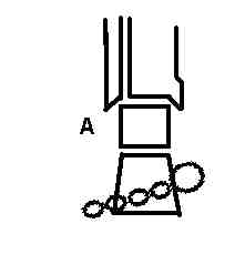
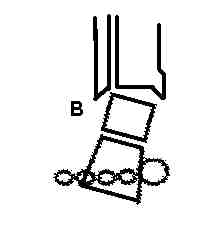
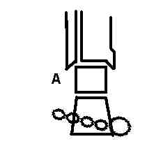
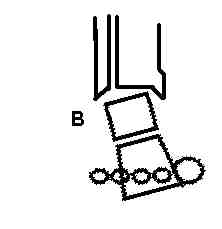
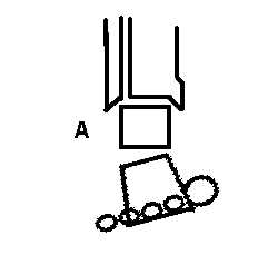
The figures illustrate four common structural variations in subtalar and foot anatomy. These structural variations result in predictable compensations. The compensations may lead to pain and dysfunction. In each case, an orthosis can support the deformity and eliminate the need for compensation (Tiberio, 1988).

Rearfoot varus

In uncompensated rearfoot varus (A), calcaneus (and forefoot) are inverted when the subtalar joint is neutral.
The person compensates (B) by pronating STJ to allow medial heel to touch the ground.
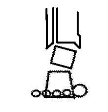
The compensation results in excessive pronation during loading response. The natural return to subtalar neutral during midstance and terminal stance is delayed.

The therapist can eliminate the need for compensation by supporting the deformity using an orthosis with a medial heel wedge.

Forefoot varus

When the subtalar joint is neutral (A), the plane of metatarsal heads is inverted in relation to the rearfoot plane.

The person compensates (B) by pronating the STJ to allow the first metatarsal to contact the ground.

The compensation results in excessive pronation during midstance, when the forefoot needs to contact the floor. This late stance pronation prevents the foot from moving to its more stable and supinated posture for pushoff. The therapist can eliminate the need for compensation by using an orthosis with a medial forefoot wedge.

Forefoot valgus

When the STJ is in neutral (A), the plane of metatarsal heads is everted in relation to the rearfoot plane.

The person compensates (B) by supinating the STJ during midstance then pronating during terminal stance.

This compensation renders the foot unstable during preswing and push-off and, over time, produces joint laxity and pain. The therapist can eliminate the need for compensation by using an orthosis with a lateral forefoot wedge.

Ankle Joint Equinus

If a person's ankle lacks normal dorsiflexion PROM when the STJ is in neutral, the person compensates by pronating the STJ during midstance and terminal stance.

This compensation provides functional dorsiflexion, but at the cost of rendering the foot mobile during push-off.

The therapist instructs the person in stretching exercises to increase isolated ankle PROM.


Resources: Michaud, T.C. (1997). The forefoot varus deformity: 9 or 90 percent prevalence? Biomechanics. [On-line]. Available: http://www.biomech.com/db_area/archives/1997/9705custom.bio.html.

Pribut, S. (1998). Biomechanics of foot and leg problems. Dr. Stephen M. Pribut's Sport Page [On-line]. Available: www.clark.net/pub/pribut/spgait.html

Tiberio, D. (1988). Pathomechanics of structural foot deformities. Physical Therapy, 68, 1840-1849.


Last updated 6-30-00 ©Dave Thompson PT
Instructors home page