Each pair of students needs a copy of:
- Hertling, D., & Kessler, R. M. (1996). Management of common musculoskeletal disorders: Physical therapy principles and methods. (3rd ed.). Philadelphia: J.B. Lippincott.
- Kendall, F.P., McCreary, E.K., & Provance, P.G. (1993). Muscles: Testing and Function (4th ed.). Baltimore: Williams & Wilkins.
- Smith, L.K., Weiss, E.L. & Lehmkuhl, L.D. (1996). Brunnstrom's Clinical Kinesiology (5th ed.). Philadelphia: F.A. Davis.
- Use your textbooks, and your knowledge of anatomy and biomechanics to answer the following questions about a person who is diagnosed with lateral epicondylitis (also called lateral tendinitis or lateral tennis elbow).
- What anatomical structures are injured?
- Of these structures, which one is most often involved and why?
- Predict two histories or scenarios, other than the ones you find in your texts, that incorporate plausible mechanisms of injury for a person who develops this problem.
- Describe two clinical tests that "rule in" the diagnosis of lateral tendinitis, or that at least increase your suspicion that the diagnosis is accurate. Devise anatomical or biomechanical explanations for both tests.
- For one of the clinical tests, describe a situation where the test will produce a "false positive" response. (A false positive response is a positive finding in a person who does not have lateral tendinitis.)
- To what other parts of the body does pain refer? Why? (Hint: examine dermatomes and sclerotomes.)
- Describe two interventions for a person with lateral epicondylitis and devise anatomical or biomechanical explanations for both. Consider issues like tissue tension and viscoelastic properties.
- Hertling and Kessler (1996, p. 185) promote "unloading" during the early mobilization of a person with shoulder impingement. Unloading is important because acutely impinged structures are so vulnerable to reinjury that "the weight of the arm alone is enough to aggravate pain" (p.186).
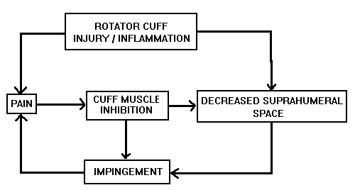
The figure (Thompson & Ahaus, 1995) illustrates how pain theoretically leads to inhibition of the rotator cuff, leading in turn to a cycle of disuse, weakness, and reimpingement.

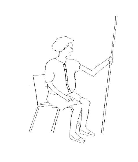
The pictured exercise employs a dowel, broomstick, or other device that bears the weight of the person's upper limb. The exercise lets one duplicate or approximate a reaching activity, but places extremely low demands on injured muscles.
With your lab partners, investigate this exercise and decide how you might use it to achieve Hertling and Kessler's (1996, p. 186) goals for "unweighted axial humeral rotation" as well as abduction and flexion. Consider:

- In what directions should the person move the dowel to achieve specific unweighted glenohumeral movements (rotation, abduction, flexion)?
- What is an appropriate trunk posture?
- How would you progress the exercise? That is, how would you increase the person's active-assistive range of motion without introducing additional weight?
- Does this approach to unweighted shoulder exercise help you think of other techniques that can strengthen extremely weak rotator cuff muscles?
Further information on the exercises
- People with rheumatoid arthritis can develop deformities of the hand when its extensor assembly (Smith, Weiss, & Lehmkuhl, 1996, pp. 198-200) is disturbed.
Use your textbooks and your knowledge of anatomy and biomechanics to answer the following questions about the "boutonniere deformity."
image of boutonniere deformity from web page of hand surgeon Charles Eaton MD.

- Describe the finger joint positions that characterize a "boutonniere deformity."
- What part of the extensor mechanism is disrupted, and consequently shifts its position to create the deformity?
- In which direction does it shift? (Hint: in the finger, flexion is produced by forces on a joint's palmar side, and extension is produced by forces on the dorsal side.) Diagram a sagittal plane view of the MP, PIP, and DIP joints to illustrate the deformity's biomechanical source.
- During exercise, muscles produce forces at joint surfaces. They also produce forces at other surfaces like fracture sites.
Use vector resolution to evaluate the effect of iliopsoas action on the site of an intertrochanteric fracture.
The solid reference line illustrates the fracture line. The vector illustrates the effect of the iliopsoas at its point of application on the lesser trochanter.
Illustrate and explain how the muscle's force produces shearing and compression (or distraction) at the fracture site.

Reference:
Thompson, D.M., & Ahaus, E.K. (1995, June). An exercise approach for older patients with impingement syndrome. Poster session presented at World Congress of Physical Therapy, Washington, DC.
Last updated 1-29-01 Dave Thompson PT
return to PHTH 7243/ OCTH 7242 lab schedule
- What anatomical structures are injured?
- Of these structures, which one is most often involved and why?
- Predict two histories or scenarios, other than the ones you find in your texts, that incorporate plausible mechanisms of injury for a person who develops this problem.
- Describe two clinical tests that "rule in" the diagnosis of lateral tendinitis, or that at least increase your suspicion that the diagnosis is accurate. Devise anatomical or biomechanical explanations for both tests.
- For one of the clinical tests, describe a situation where the test will produce a "false positive" response. (A false positive response is a positive finding in a person who does not have lateral tendinitis.)
- To what other parts of the body does pain refer? Why? (Hint: examine dermatomes and sclerotomes.)
- Describe two interventions for a person with lateral epicondylitis and devise anatomical or biomechanical explanations for both. Consider issues like tissue tension and viscoelastic properties.
The figure (Thompson & Ahaus, 1995) illustrates how pain theoretically leads to inhibition of the rotator cuff, leading in turn to a cycle of disuse, weakness, and reimpingement.

|
|---|
The pictured exercise employs a dowel, broomstick, or other device that bears the weight of the person's upper limb. The exercise lets one duplicate or approximate a reaching activity, but places extremely low demands on injured muscles.With your lab partners, investigate this exercise and decide how you might use it to achieve Hertling and Kessler's (1996, p. 186) goals for "unweighted axial humeral rotation" as well as abduction and flexion. Consider: | 
|
|---|
- In what directions should the person move the dowel to achieve specific unweighted glenohumeral movements (rotation, abduction, flexion)?
- What is an appropriate trunk posture?
- How would you progress the exercise? That is, how would you increase the person's active-assistive range of motion without introducing additional weight?
- Does this approach to unweighted shoulder exercise help you think of other techniques that can strengthen extremely weak rotator cuff muscles?
Further information on the exercises
Use your textbooks and your knowledge of anatomy and biomechanics to answer the following questions about the "boutonniere deformity."image of boutonniere deformity from web page of hand surgeon Charles Eaton MD. | 
|
|---|
- Describe the finger joint positions that characterize a "boutonniere deformity."
- What part of the extensor mechanism is disrupted, and consequently shifts its position to create the deformity?
- In which direction does it shift? (Hint: in the finger, flexion is produced by forces on a joint's palmar side, and extension is produced by forces on the dorsal side.) Diagram a sagittal plane view of the MP, PIP, and DIP joints to illustrate the deformity's biomechanical source.
Use vector resolution to evaluate the effect of iliopsoas action on the site of an intertrochanteric fracture.The solid reference line illustrates the fracture line. The vector illustrates the effect of the iliopsoas at its point of application on the lesser trochanter. Illustrate and explain how the muscle's force produces shearing and compression (or distraction) at the fracture site. | 
|
|---|