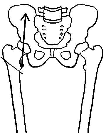
The muscle's force vector (TM) is illustrated, along with a reference line that represents the glenohumeral joint's surface
The next step involves moving the muscle's vector, without tipping it or changing its orientation, so that the vector's point of application rests somewhere the reference line.
Resolving the muscle vector involves drawing two component vectors:
- one that is parallel to (directly on) the reference line
- one that is perpendicular to the reference line.
The two vectors are called components of the original vector because, when added using the technique of vector composition, their resultant or sum is the original muscle vector.