Biomechanics of the wrist and hand

Each lab group needs these resources:
These lab problems are designed to help students review hand anatomy, to relate their existing knowledge to functional considerations, and to introduce new material.

Students should use lab time to consider the first set of problems, to ask questions, and to have their knowledge challenged by instructors and classmates.

  1. Use Smith, Weiss, and Lehmkuhl (1996, pp. 182-183) and Hertling and Kessler (1996, pp. 253-254) to review the wrist's surface anatomy and to palpate the wrist's muscles, tendons, ligaments, and its eight bones.

    As you review wrist anatomy, note:

    • the carpal tunnel, through which the median nerve passes. Therapists employ a special maneuver, Phalen's test, to detect median nerve entrapment or compression in the carpal tunnel. In one study (Gellman, Gelberman, Tan, & Botte, 1986), Phalen's test (Hertling & Kessler, 1996, Fig. 17-25, pp. 544,567) detected median nerve involvement with specificity and sensitivity of 0.71 and 0.50, respectively. Draw on your knowledge of anatomy and function to explain the rationale for Phalen's test. Ponder why it sometimes yields false positive and false negative results.

    • the tunnel of Guyon (Hertling & Kessler, 1996, p.254), through which pass the ulnar artery and ulnar nerve.

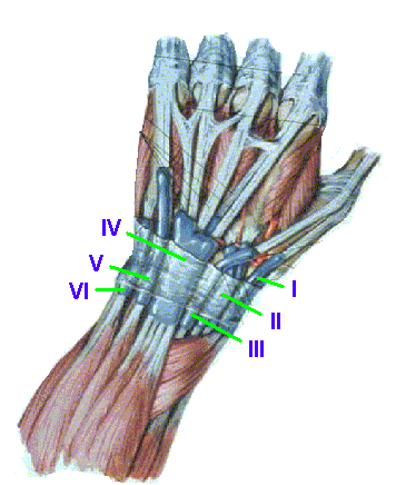
    • the dorsal tendon compartments

      Identify the tendons that, encased in synovial tendon sheaths, comprise six numbered dorsal compartments of interests to hand therapists:

      1. abductor pollicis longus and extensor pollicis brevis
      2. extensor carpi radialis longus and brevis
      3. extensor pollicis longus
      4. extensor digitorum comunis (four tendons) and extensor indicis
      5. extensor digiti minimi
      6. extensor carpi ulnaris

      5 dorsal tendon compartments

  2. Opening of the hand

    To understand the muscular synergies involved in opening the hand (and in the next problem, which involves closing the hand), you should examine the extensor mechanism and the muscles that attach to it. Refer to a popular anatomy atlas like Netter (1997, Plate 433 - Flexor and extensor tendons in fingers), to your text's reproductions of Netter's drawings of the extensor mechanism (Smith, Weiss, & Lehmkuhl, 1996, Fig 6-12), or to adaptations of Netter's drawings.

    The extensor digitorum "is mechanically capable of extending the MCP, PIP, and DIP joints but not at the same time. When the extensor digitorum contracts alone, ... the MCP joints extend but the IP joints remain semiflexed in a clawhand position (Smith, Weiss, & Lehmkuhl, 1996, p.205-6)." The authors also explain that the extensor digitorum combines with the lumbricales in a muscle synergy to open the hand. Unless the hand must be opened forcefully or against resistance, the interosseous muscles are inactive (pp. 206-207).

    1. How can the lumbricales contribute to hand opening?

    2. How can the interossei contribute to forceful opening of the hand?

  3. Forceful closing of the hand

    According to Smith, Weiss, and Lehmkuhl (1996, p. 201), "forceful closure of the hand or power grip elicits high-level activity of the flexor digitorum superficialis, the interossei, and the flexor digitorum profundus."

    Explain why we use the interosseous muscles in hand closure, even though they can contribute to PIP and DIP extension.

  4. Review the saddle-like structure and biaxial function of the first carpo-metacarpal joint.

    The thumb's CMC joint abducts and adducts in a plane perpendicular to the palm. Some therapists specify the movement toward abduction as "palmar abduction."

    The first CMC joint also flexes and extends in a plane that is parallel to the palm. Some therapists refer to extension in this plane as "radial abduction."

    Opposition and its antagonistic movement, reposition, involve an automatic rotation of the first metacarpal that occurs when certain movements are combined. You can verify this if you:

    abduct, then flex the first CMC: the metacarpal will rotate as the CMC joint moves into opposition.

    extend and adduct the first CMC: the metacarpal rotates the opposite direction as the joint moves toward reposition.

    Note that you cannot passively rotate the CMC after you have placed it in full opposition or full reposition. Those are the joint's close packed positions. The joint's capsular fibers are elongated as the joint approaches either end of its range of motion. Once they capsule is maximally elongated and its ligamentous fibers are maximally, the surfaces cannot rotate further.

  5. Types of grasp Therapists classify grasp or prehension patterns as power grasps or precision grasps according to the position and mobility of the thumb's CMC and MP joints. Observe your hand while you perform the prehension patterns listed in your texts (Hertling & Kessler, 1996, 259-260; Smith, Weiss, & Lehmkuhl, 1996, pp.216-218) and verify that the different grasps require different positioning and movement in the thumb and digits.

Students should consider the second set of problems during the lab session. However, the problems contain enough background information for students to work on them independently after lab.

  1. Synergies among thumb muscles

    • Rapid flexion of the thumb's IP joint involves the flexor pollicis longus (FPL), and is often accompanied by palpable activity in the abductor pollicis longus. The first CMC joint may even abduct. The APL activity counters FPL's tendency to flex the CMC joint in the plane of the palm.

    • Rapid extension of the thumb's IP joint involves the extensor pollicis longus (EPL), and is often accompanied by palpable activity in the thenar muscles. The first CMC joint may even flex in the plane of the palm. Activity in the opponens pollicis counters the EPL's tendency to extend the first CMC joint or adduct it in a plane perpendicular to the palm.

    • The abductor pollicis longus (the APL, a snuffbox muscle) acts in synchrony with any action of the thenar muscles. Its activity stabilizes the first metacarpal so that the thenar muscles do not produce movement at the first CMC joint. Since the APL also produces radial deviation of the wrist, the extensor carpi ulnaris (ECU) may also act to prevent wrist motion.

  2. Test for motor integrity of the anterior interosseous nerve

    Special tests are designed around knowledge of anatomy (including innervation) and function. Hertling and Kessler (1996, p. 261) describe a specific functional deficit in a person whose anterior interosseous nerve is compressed. "During pinch the distal phalanges of the thumb and index finger cannot flex and stay in extension."

    • The anterior interosseous nerve is a branch of the _____ nerve.

    • The person cannot flex the index finger's DIP because of weakness in the ____________.

    • The person cannot flex the thumb's IP joint because of weakness in the _____________.

    • To hold an object, the person compensates by permitting the joints to extend, and uses muscles that innervated by the _______ nerve.

  3. Froment's sign

    Froment's sign (Rothstein, Roy, & Wolf, 1991, pp. 126-7) is the eponym for a special test that assesses function in a specific nerve. The subject performs the test by grasping a piece of paper between the tip of the thumb (with the IP joint extended) and the radial side of the second digit. Froment's sign is judged to be positive if the person must flex the thumb's IP joint to maintain a grasp when the examiner attempts to pull the paper from the person's fingers.

    When the examiner notes a positive Froment's sign, he or she judges that:

    1. the patient must use the _________________ (extrinsic thumb muscle that flexes the IP joint),

    2. which is innervated by the _________________________ nerve

    3. to compensate for weakness in the _______________________ (intrinsic thumb muscle),

    4. which is innervated by the ______________________ nerve.

    5. The test detects problems with the ___________ nerve.

Students who have additional time on their hands can address the third set of problems.

  1. Contribution of the extrinsic thumb extensors to adduction of the first MCP joint.

    The extensor pollicis longus and brevis "extend the thumb and from this position can act as [MCP] adductors" (Smith, Weiss, & Lehmkuhl, 1996, p. 211). Explain this observation in terms of the muscles' lines of application.

  2. Differences in the attachments of the interosseous muscles

    Resist the action of each of your partner's interosseous muscles in abduction or adduction of the appropriate MCP joint. In many people, abduction is more forceful in the second digit than the others.

    1. How does the interosseous muscles' anatomy account for this difference?

    2. What implications might this have for hand function?

References:

Gellman, H., Gelberman, R.H., Tan, A.M., & Botte, M.J. (1986). Carpal tunnel syndrome: An evaluation of the provocative diagnostic tests. Journal of Bone and Joint Surgery, 68A, 735-737.

Rothstein, J.M., Roy, S.H., & Wolf, S.L. (1991). The rehabilitation specialist's handbook. Philadelphia: F.A. Davis.


Last updated 3-15-01 Dave Thompson PT
return to PHTH 7243/ OCTH 7242 lab schedule