Case No.: N-017 Quiz
Diagnosis: Anaplastic oligodendroglioma, WHO grade III with chromosome 1p and 19q deletion
Organ: Brain, cerebral hemisphere
Last Updated: 09/21/2010
|
|
|
Hematoxylin & eosin |
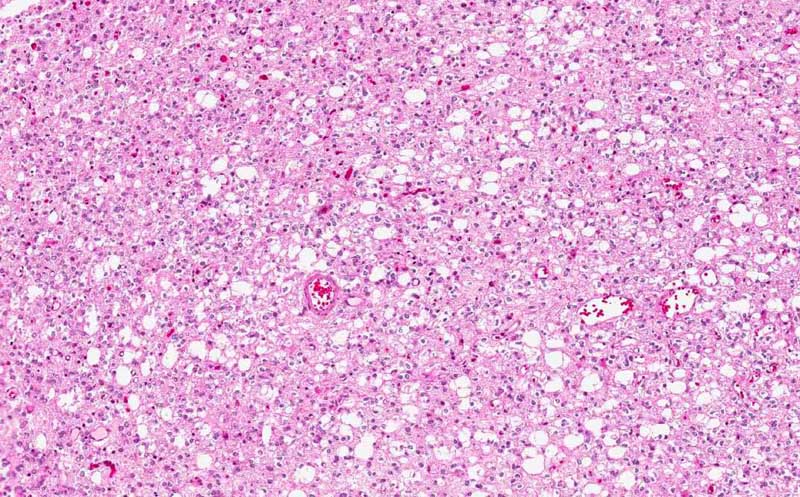
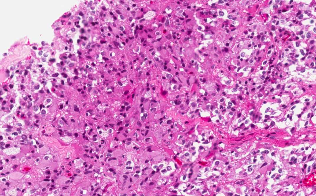
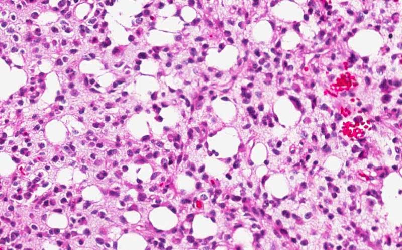
Area 1: In this this area, the tumor cells are solidly packed. The characteristic feature of "fried eggs" appearance featured by round to polygonal cells, prominent perinuclear halo, round and centrally located nuclei are diagnostic features of oligodendroglioma. There are also many small cyst in this area. Note that there is a vascular network composed of thin capillaries (arrow) embedded within the tumor. |
|
Hematoxylin & eosin |
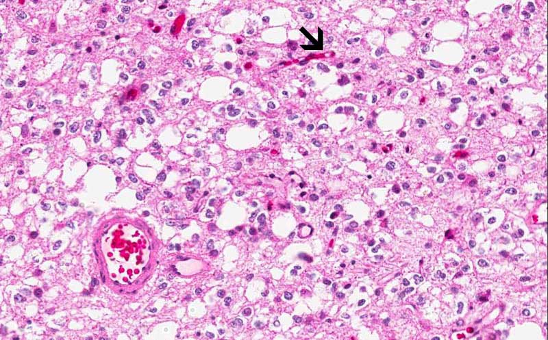
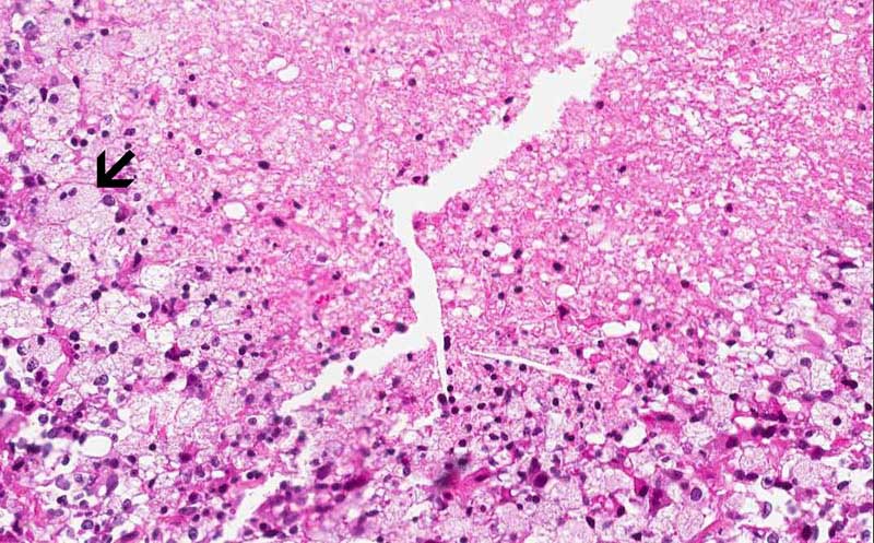
Area 2: This is an area of gray matter infiltrated by the tumor. Note that the tumor cells are not solidly packed back-to-back. Rather, there is brain parenchyma between tumor cells. Many amorphous calcifications (white arrow) are present. There are also some neurons entrapped between the tumor cells (black arrow) |
|
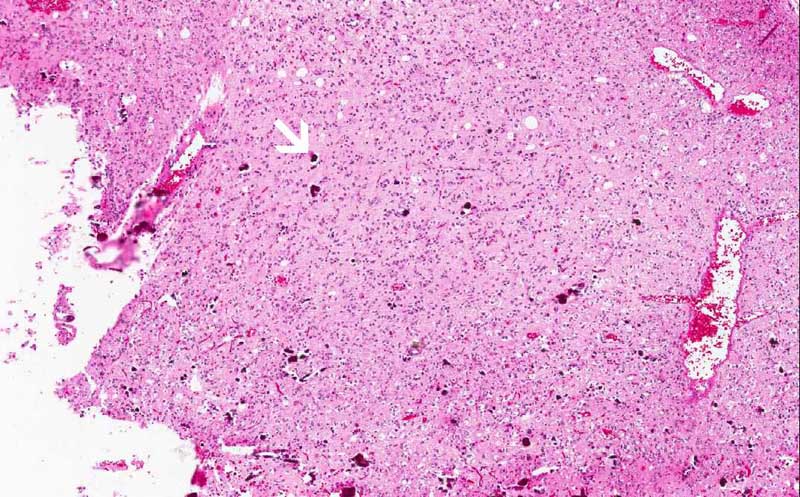
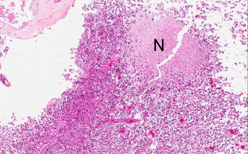
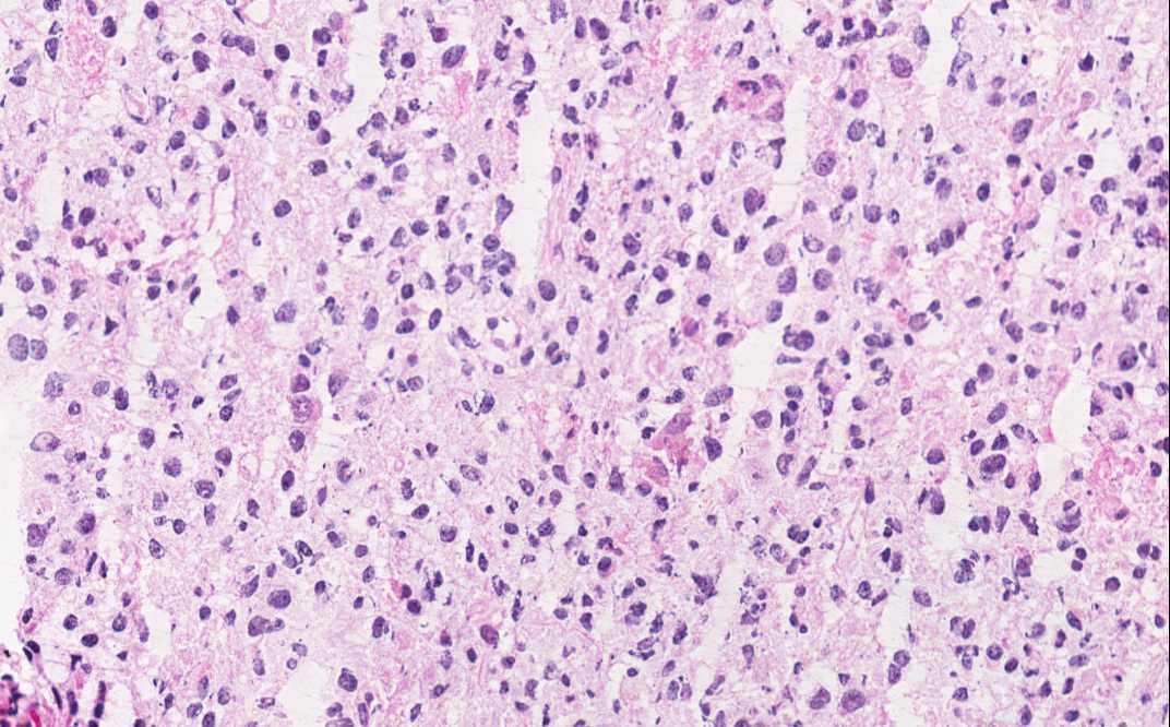
Hematoxylin & eosin Area 3: There is a small area of necrosis in this area (N). Note that some of the cells have amphophilic cytoplasm rather than clear perinuclear halo. There is also increased pleomorphism in these cells. When oligodendroglioma cells with increased atypia, their cytoplasm often lost the clear halo and there is also increase in variation of nuclear size and diameter. Note that there are some foamy cells with small nuclei around the necrotic debris. These are foamy histiocytes (macrophages) and should not be mistaken as tumor cells (arrow). |
|
Hematoxylin & eosin |
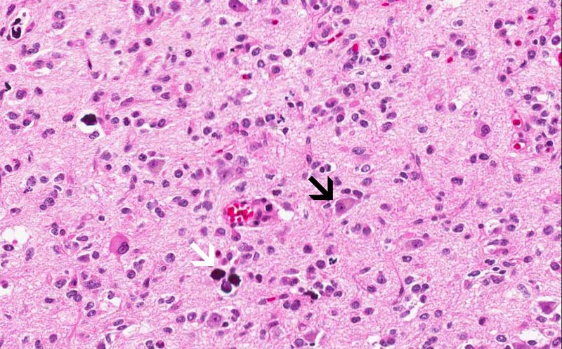
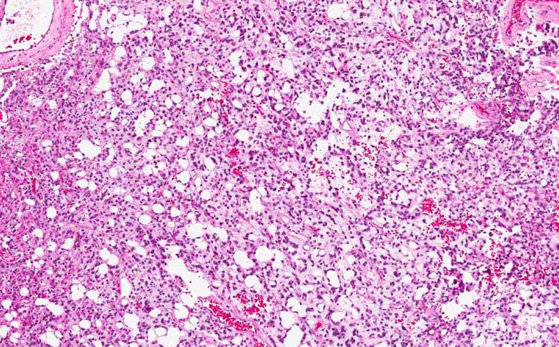
Area 4: In this area, there is substantial microcyst formation which is very common in oligodendroglioma. The tumor cells appear less "fried egg" like than than other areas. |
|
History: The patient was a 42 year-old woman who developed seizure at home and was taken to the emergency room by her family members. On further questioning, she also has a history of chronic headache for a few weeks. Fundic examination demonstrated papillary edema of the optic cup. An MRI scan demonstrated a 3 cm enhancing mass in the frontal lobe associated with substantial edema and midline shift. The tumor was excised and yielded the specimen being shown here.
Histologic Highlights of this Case:
Comment:
|
Bonus Images:
|
Hematoxylin & eosin |
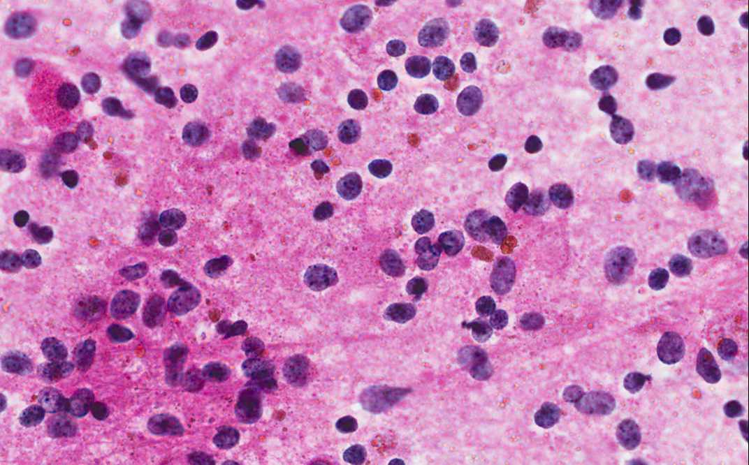
Intraoperative Cytologic Preparation: Note that the tumor cells are mostly in form of naked nuclei with a monotonous, rather round morphology. |
|
Hematoxylin & eosin |
Intraoperative Frozen Section: Note that the perinuclear halo is a formalin fixation artifact and is not present in frozen sections. |
Original slide is contributed by Dr. Kar-Ming Fung, University of Oklahoma Health Sciences Center, Oklahoma, U.S.A.