Answer and Discussion of Quiz Set: N-019
6. Which of the following features will be helpful in the diagnosis and prognostic assessment of the tumor illustrated below which is obtained from the frontal lobe of a 32 year-old man? Answer

A. Positive immunoreactivity for glial fibrillary acidic protein (GFAP)
B. Mutation of isocitrate dehydrogenase (IDH) 1 and lost of chromosome 1p and 19q
C. Weakly positive for synaptophysin staining
D. Demonstration of BRAF-KIAA1549 fusion
E. Demonstration of gemistocytic astrocytes
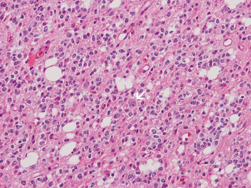
Answer and Discussion: The answer is (B). The tumor being shown here is most consistent with an oligodendroglial tumor. The image being shown here is obtained form an oligodendroglioma, WHO grade II. characterized by delicate blood vessel network, rather monotonous and round nuclei, and perinuclear halo. Note that there is an entrapped neuron (arrow) at the center of the lesion. This is a testimony to the invasive nature of this tumor. These tumors often have mutation of IDH. A lost of chromosome 1p and 19q can be seen in some of the cases and indicate responsiveness to chemotherapy.
Oligodendroglial tumors can be positive for GFAP and can have minigemistocytes. Both of them are not diagnostic, however. Some of them also have positivity for synaptophysin. However, when the immunoreactivity for synaptophysin is strong and that the tumor is close to the ventricles, a possibility of central neurocytoma should be considered.
Pilocytic astrocymoa often have BRAF-KIAA1549 fusion. Some of them may also have nuclear halos which raise the suspicion of an oligodendroglioma. However, the age and location of this tumor does not suggest pilocytic astrocytoma. Also, there is no classic histologic features suggestive of a pilocytic astrocytoma here. Also, the entrapped neurons within tumor cells is not a common feature of pilocytic astrocytoma which is non-invasive and well demarcated in most cases.