Case No.: N-027 Quiz
Diagnosis: Chordoid meningioma, WHO grade II
Organ: Brain
Last Updated: 11/21/2011
|
|
|
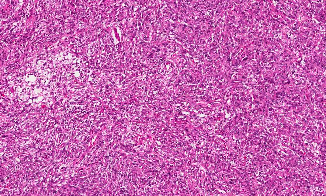
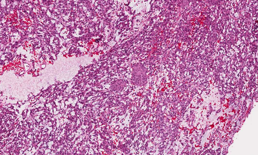
Hematoxylin & eosin |
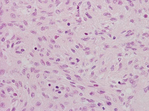
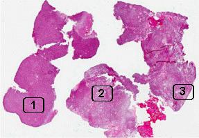
Area 1: The tumor cells are rather spindle in shape and have a vague concentric arrangement. However, classic meningotheliomatous whorls are not present. This type of vague concentric arrangement of tumor cells is a hint for diagnosis in meningiomas. If you pay attention, the cytoplasm of the tumor cells have a fine bubbly type of appearance. |
|
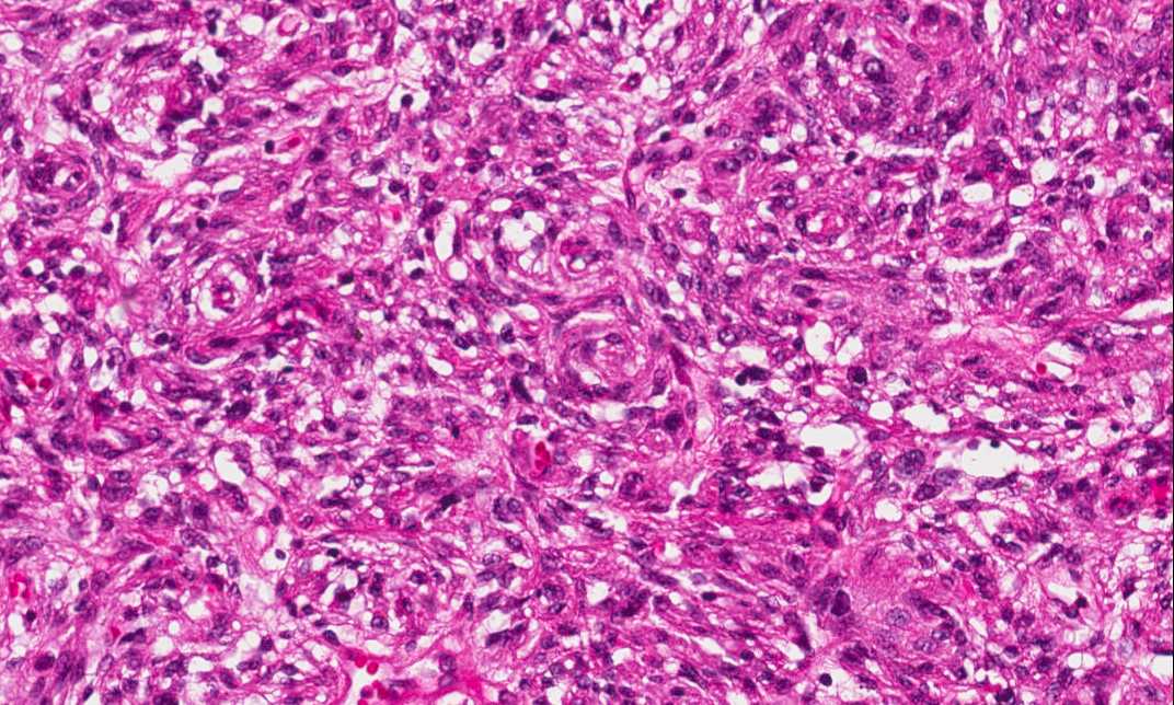
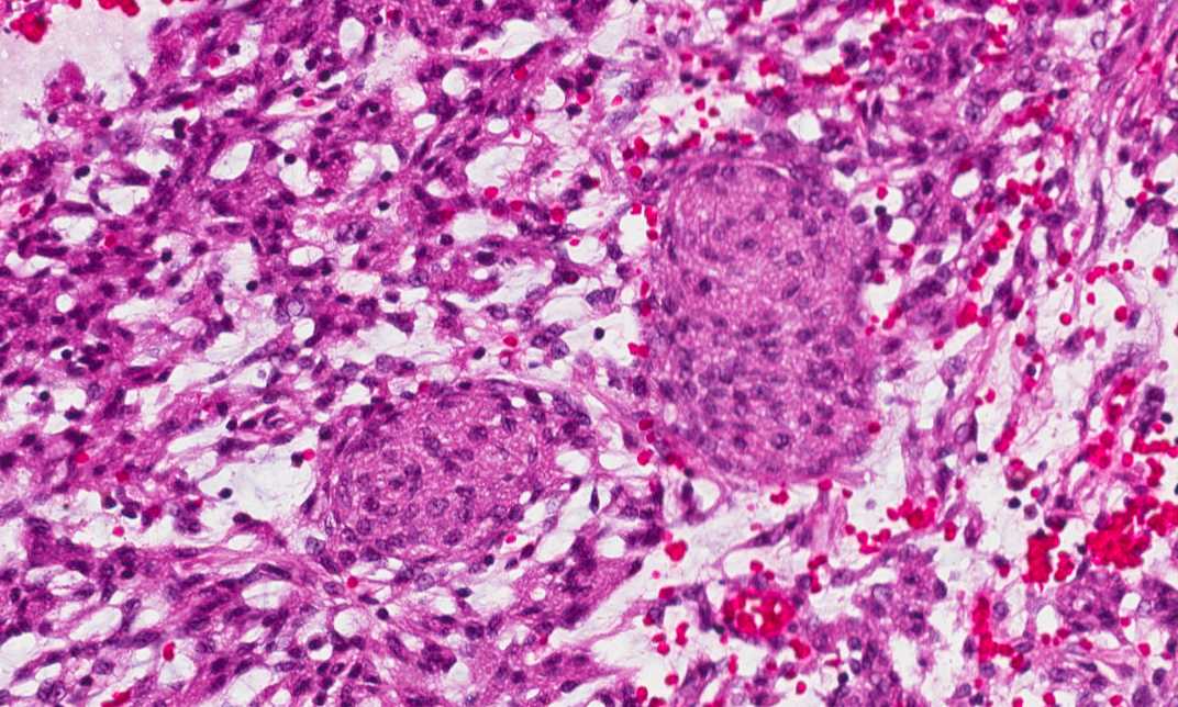
Hematoxylin & eosin |
Area 2: This area has substantial amount of intercellular mucoid material in between tumor cells. It looks quite different from Area 1. However, if you put in some imagination and mentally add the intercellular mucoid material to Area 1, the two areas do not look that much different anymore. |
|
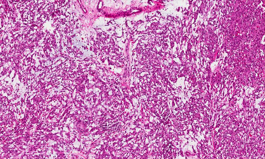
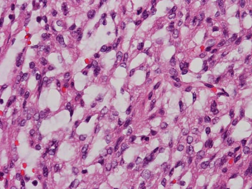
Hematoxylin & eosin |
Area 3: In this area, there are some islands that are suggestive of meninogthelial meningioma component. If you pay attention, however, these cells also have fine bubbly cytoplasm. |
|
History: The patient was a 56 year-old woman who presented to his primary care physician with a history of headache for 3 months. An MRI scan demonstrated an enhancing 3 cm dural based mass in the frontal lobe.
Histologic Highlights of this Case:
Comment:
|
Bonus Images:
|
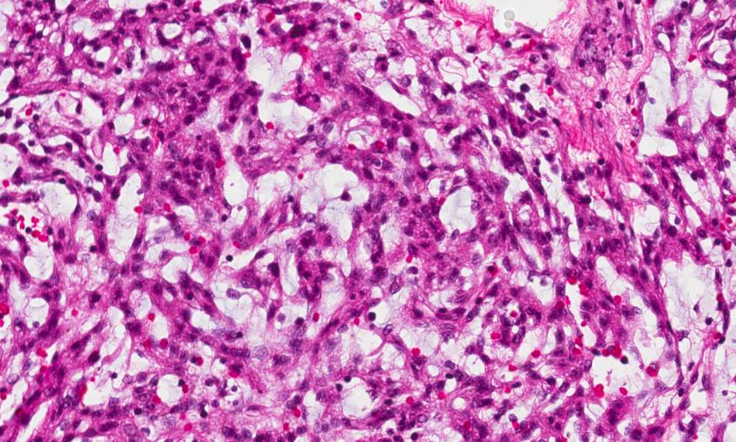
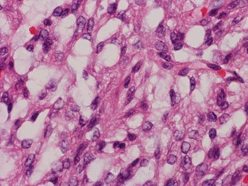
Hematoxylin & eosin |
High Magnification: The fine bubbly cytoplasm is similar to the physaliferous (Greek for "bubble-bearing") cells in chordoma. For this features and the mucoid intercellular substance, these tumor are named chordoid meningioma. |
|
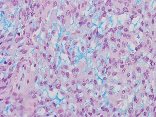
Hematoxylin & eosin |
Alcian blue stain: The Alcian blue positive intercellular substance is not present in the more solid area. In areas that are less solid area, the mucoid intercellular substance is strongly positive for Alcian blue. |
Original slide is contributed by Dr. Kar-Ming Fung, University of Oklahoma Health Science Center, Oklahoma, U.S.A.