Case No.: W-001   Quiz 

Diagnosis: Metastatic adenocarcinoma, lung origin   

Organ: Vertebral column, L2

Last Updated: 12/22/2009


Online Slide/Full Screen/

Open with ImageScope

CT: The osteolytic lesion is highlighted by the arrow.
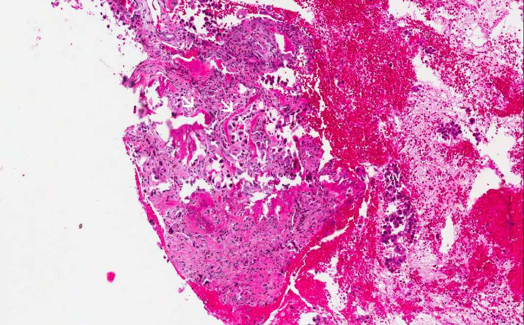
Hematoxylin & eosin

Area 1: Under low-magnification, small fragments of bone (arrow) surrounded by atypical cells are present in this biopsy specimen.
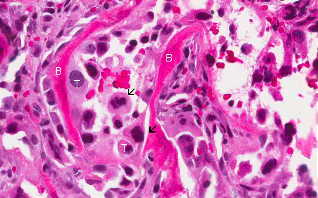
Hematoxylin & eosin

Area 1: The bone fragments (B) are surrounded by large, pleomorphic, epithelial cells (T) that are diagnostic morphologically for a metastatic non-small cell carcinoma. Many mitotic figures are present (arrows). Note that some of the cells around the bone chips has elongated or a spindle appearance.
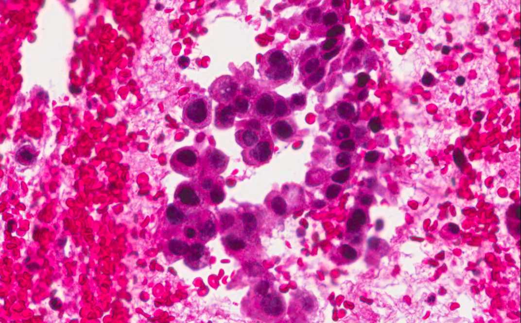
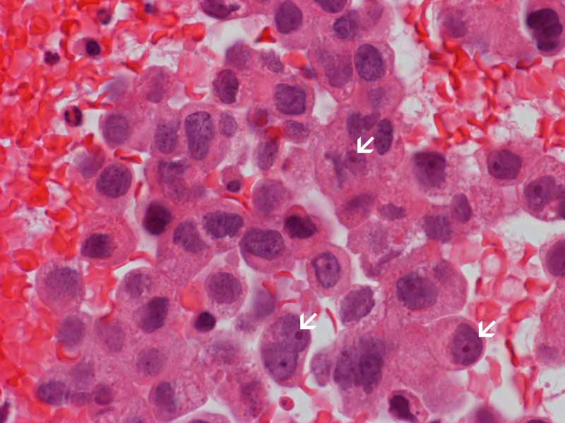
Hematoxylin & eosin

Area 1: In the same specimen, there are clusters of pleomorphic epithelial cells not attached to the bone. In these areas, the epithelial nature of a carcinoma is most clearly seen.

History: The patient was a 58 year-old woman with low back pain for about 5 months duration. There was no history of fall, trauma, or other events that precipitated the pain. The patient was a house wife and smoke about 1 pack of cigarettes a day but with denied the use of alcohol or drug. On CT imaging, an osteolytic lesion was demonstrated in the posterior elements of her L2 spine. A needle biopsy was performed and yielded the the current specimen.


Histologic Highlights of this Case:

  • Due to the presence of bone fragments, the specimen was decalcified with acid after fixation with formalin for histological sections.

  • The specimen is composed mainly of blood. In some small areas of the specimen, there are bone fragments surrounded by large, atypical, epithelial cells. The morphology is most consistent with a metastatic non-small cell carcinoma.

  • Morphology alone cannot always determine the origin of a metastatic tumor. Immunohistochemistry was performed in this case. The overall profile is as follow:

  • Pancytokeratin, cytokeratin 7, thyroid transcription factor 1 (TTF-1): positive in tumor cells.

  • Cytokeratin 20: Negative in tumor cells.

  • Gross cystic disease fluid protein 15 (GCDFP-15): Negative in tumor cells.

  • This profile suggest that the tumor is most likely a metastatic adenocarcinoma of lung origin. The negative GCDFP15 result rule out the possibility of metastatic breast carcinoma in this case.

Bonus Images:

Hematoxylin & eosin

Additional high-magnification images: Note the relationship between the bone (B) and tumor cells (arrows).

Hematoxylin & eosin

Additional high-magnification images: Note the enlarged nuclei with nucleoli (arrow).

Immunohistochemistry

Pancytokeratin (AE1/AE3): Note the positive cells (brown cells) and bone (B). Most carcinomas are positive for this combination of cytokeratin on immunohistochemistry.

Immunohistochemistry

Cytokeratin 7:  Cytokeratin 7 is often but not always positive in adenocarcinoma and small cell carcinoma arising from the lung. In contrast, cytokeratin 20 is often negative in these tumors.

Immunohistochemistry

Thyroid transcription factor 1 (TTF-1):  TTF-1 is often positive in adenocarcinoma and small cell carcinoma of the lung. They are also positive in tumors of the thyroid gland. In contrast to the staining in AE1/AE3 and cytokeratin 7, the positive staining in TTF-1 is in the nuclei.

Original slide is contributed by Dr. Kar-Ming Fung, University of Oklahoma Health Sciences Center, Oklahoma, U.S.A.

 Home Page