
This is a translational research project funded by the Mary Kay Ash Charitable Foundation to identify proteins that can be used to predict the tumorigenic behavior and retinoid sensitivity of ovarian cancer cells. In this project, ovarian cell lines and primary culture of normal and neoplastic ovarian tissue are grown inside as well as on top of the collagen. Histological evaluation of the cultures revealed the presence of glands and mucin expression that was increased by retinoids in some culture. Although normal ovarian cells do not express mucin, it's expression is a characteristic of low grade tumors. We have demonstrated that some retinoids induce gland formation and mucin expression in organotypic cultures of ovarian cancer cells but not of normal ovarian cells. Therefore it appears that retinoids are reversing tumorigenesis to an earlier stage.
 |
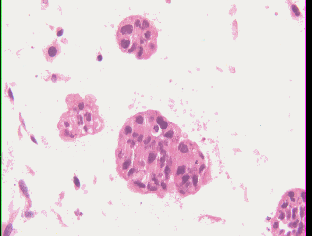
Caov-3 Ovarian Cancer Cell Line Grown in Organotypic Culture in the absence of retinoids. The malignant cells are present both as scattered single cells and as clumps inside the collagen. The clumps are composed of cells arranged chaotically. Crowding is present. |
 |
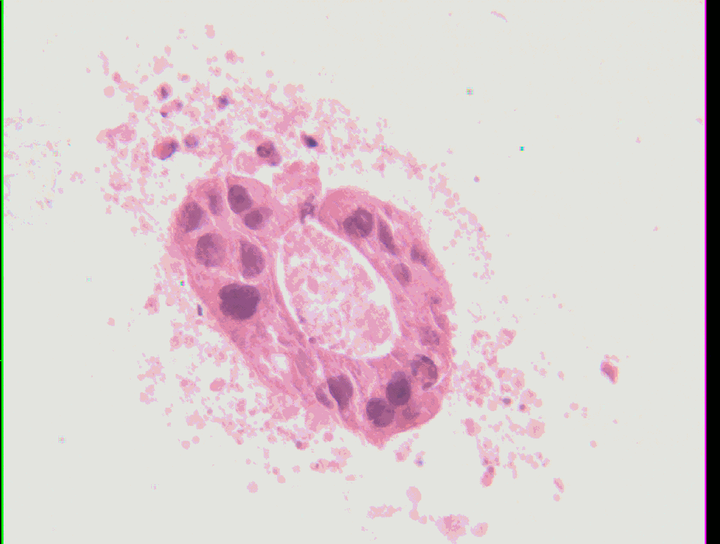
Caov-3 organotypic cultures treated with SL173 retinoid. In contrast to the control, the majority of clumps now have central areas containing debri and some apoptotic cells. Rare well-formed glands are present. Where gland formation is present, the cells are arranged around a central space with the individual cells that have cytoplasm pointed toward the space and their nuclei located at the outer periphery of the cells. |
Eleven different retinoids were evaluated for their effects in this organotypic culture model system of ovarian tumorigenesis. Some of the retinoids induced a natural form of cell death, called apoptosis, of the ovarian tumor cells (detected by the Tunnel Assay), some induced differentiation (detected by observation of gland formation and positive PAS staining), and some induced both apoptosis and differentiation. The extent of apoptosis strongly correlated with the degree of growth inhibition, but the extent of differentiation did not.
The second year of the project will include an animal model to test the best retinoids identified in the first year of the project and include an evaluation of biomarker expression in the organotypic cultures that were generated during the first year of this project. To further explore the role of apoptosis verses differentiation in the mechanism of retinoid activity against ovarian tumor cells, the xenograft animal model planned for the second year of this project will compare retinoids that exhibit different apoptosis and differentiation activities.
The observation of mucin induction by retinoids has lead to a review of the literature in order to understand the role of mucin expression in ovarian cancer. This literature review has lead to more questions than answers that are addressed in a NIH grant proposal in preparation. Therefore this project has generated significant findings and future directions that provide promise for developing improved care for women with ovarian cancer.