Muscles produce forces. Because the forces act at a distance from a joint's axis of rotation, they also produce moments at the joint. Muscle forces simultaneously influence arthrokinematics, movement at the joint's surface. We can evaluate the forces' effects on arthrokinematics using the technique of vector resolution.
- Verify that the middle deltoid produces abduction around the glenohumeral joint's A-P axis. Verify additionally that the middle deltoid causes superior shearing or sliding of the humeral head on the glenoid fossa.
- Verify that the infraspinatus has a very small moment with respect to the glenohumeral joint's A-P axis and, therefore, has minimal effect on glenohumeral abduction. However, the infraspinatus is an important synergist in glenohumeral abduction because of its effect on arthrokinematics. Verify that the infraspinatus causes inferior shearing or sliding of the humeral head on the glenoid fossa.
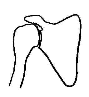
To verify each muscle's effect on shearing or sliding, perform vector resolution using the diagrams below (one to illustrate the middle deltoid, the other to illustrate infraspinatus):

|

|
|---|
for the middle deltoid for the infraspinatus
|
|---|
The force component whose vector is perpendicular to the reference line represents that part of the muscle's force that causes the humerus to ______ the glenoid fossa.
for the middle deltoid for the infraspinatus
|
|---|
Next semester, you will extend your knowledge of manual muscle testing to the upper limb. Preview this skill, and reinforce your anatomical knowledge by following your text's (Kendall, McCreary, & Provance, 1993) directions to test the strength of:
- any two parts of the trapezius
- one other scapulothoracic muscle
- one rotator cuff muscle
- one part of the deltoid muscle
Smith, Weiss, and Lehmkuhl (1996, p. 235) remark that "90 to 100 degrees of motion occur at the glenohumeral joint" during shoulder elevation. Verify their further observation that "achievement of the glenohumeral range requires external rotation for the shoulder in abduction and internal rotation for flexion." To assess the direction of glenohumeral rotation during abduction or flexion, palpate the epicondyles at the humerus' distal end.
Smith, Weiss, and Lehmkuhl (1996, p. 230) observe that "the combined effect of acromioclavicular and sternoclavicular motions is to permit scapular movement so that the glenoid fossa may face" in a direction that is appropriate for a task. They observe that "the sum of the ranges of motion at the sternoclavicular [SC] and acromioclavicular [AC] joints equals the range of motion of the scapula" (p.230). Of the scapular upward rotation that must occur during shoulder elevation, they attribute 30 to 45 degrees to SC joint motion (p.229) and 20 degrees to AC joint motion (p. 230).
Review the sequence of movements that occurs in the scapulothoracic, AC, and SC joints, and in the thoracic spine (Smith, Weiss, & Lehmkuhl, 1996, p. 235) during shoulder elevation. Use the laboratory skeletons to locate and appreciate the significance of the ligaments (costoclavicular and coracoclavicular) in which passive force develops and which contribute to the sequence of movements.
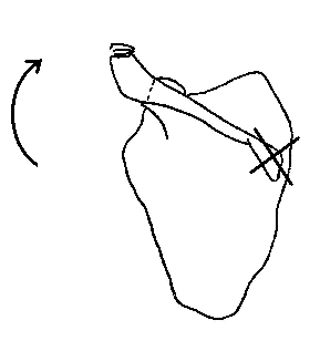
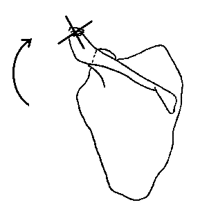
Review and diagram two important muscular synergies that produce scapular upward rotation as the shoulder elevates.

| 
|
|---|---|
Serratus anterior and upper trapezius, during the initial phase of shoulder elevation, when the scapula rotates upward around an AP axis that projects through the sternoclavicular joint to the base of the scapular spine.
Serratus anterior and lower trapezius, during a later initial phase of shoulder elevation, when the scapula rotates upward around an AP axis that passes through the acromioclavicular (AC) joint. |
We have focused most of our discussion thus far on the muscles involved in shoulder elevation. People also employ the shoulder girdle in activities that require shoulder depression. Bearing weight through the handles of a walker to achieve a non-weightbearing gait pattern is one example. Another example is the seated push-up, a function that is important to activities like a "sliding board transfer." Both of these examples involve closed chain movements wherein thorax moves while the scapula and humerus are the relatively stable segments.
Focus on the scapulothoracic joint, and analyze scapular depression during either of these activities. Remember that the movements are closed chain movements, and that the scapula is relatively stable. Understand that the moving segment is the thorax, then determine gravity's effect on it. Finally, decide which muscles act to produce the thoracic movement.
When you've given the problem some thought, you can refer to a commentary.