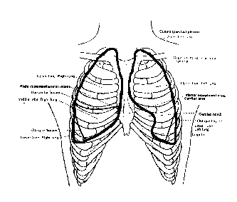
The T10 spinous process is a posterior surface landmark for the inferior boundary of lung substance (Clemente, 1981, Fig.129).
- base of scapular spine is at T4
- inferior angle of scapula is at T8
PV/nT = R
(where R is the 'gas constant')
Implications:

| The T10 spinous process is a posterior surface landmark for the inferior boundary of lung substance (Clemente, 1981, Fig.129).
|
|---|

| The xiphoid process is an anterior surface landmark for the inferior boundary of lung substance (Clemente, 1981, Fig.128). |
|---|
INSPIRATIONEXPIRATION
DIAPHRAGM DESCENDSRIBCAGE ELEVATES AND/OR EXPANDS INCREASED INTRATHORACIC VOLUME DECREASED INTRATHORACIC PRESSURE 'HIGH PRESSURE' EXTERIOR AIR FLOWS INTO 'LOW PRESSURE' LUNG. DIAPHRAGM ASCENDSRIBCAGE DESCENDS AND/OR CONTRACTS DECREASED INTRATHORACIC VOLUME INCREASED INTRATHORACIC PRESSURE 'HIGH PRESSURE' AIR IN LUNG FLOWS OUT TOWARD 'LOW PRESSURE' EXTERIOR. |
|---|
InspirationExpiration
Quiet |
elastic recoil of lung tissue
| Forced |
|---|
Diagrams of secondary (accessory) muscles of respiration
Anterior view of diaphragm (Rasch & Burke, 1974, Fig.14-3) showing its lines of application where it attaches to the ribs and to the central tendon."Top down" view of diaphragm (Clemente, 1981, Fig.193) showing location of central tendon. |
|---|
During inspiration, the diaphragm's central tendon descends until it is fixed or stabilized by forces that develop in:
When the central tendon becomes stable, it is still superior to the diaphragm's mobile attachments on the lower ribs. Therefore, the diaphragm's muscular lines of application elevate the lower ribs. Because of the orientation of the lower ribs' attachments to the vertebrae, rib elevation expands the thorax' lateral dimensions.
Clemente, C.D. (1981). Anatomy. (2nd ed.). Baltimore: Urban and Schwarzenberg.
Kapandji, I.A. (1974). Functional components of the vertebral column. In I.A. Kapandji, The physiology of the joints: Vol. 3. The trunk and the vertebral column. New York: Churchill Livingstone.
Poole, D.C., Sexton, W.L., Farkas, G.A., Powers, S.K., Reid, M.B. (1997). Diaphragm structure and function in health and disease. Medicine and Science in Sports and Exercise, 29, 738-54. (full text version is available on Medline; Unique Identifier: 97362737).
Rasch, P.J., & Burke, R.K. (1978). Kinesiology and applied anatomy (6th ed.). Philadelphia: Lea and Febiger.