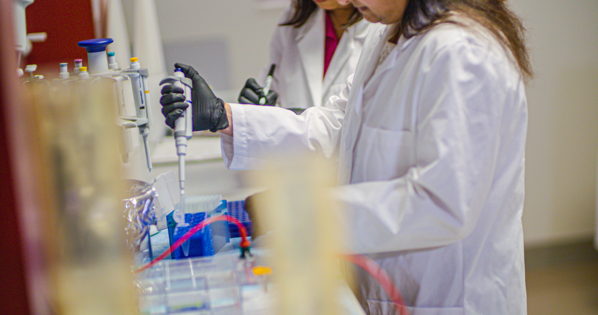
Harold Hamm Diabetes Center offers access to core facilities at a free or discounted rate for the further development of research projects. Current core facilities include the Diabetic Animal Core and Histology Core. To apply for access, please contact the core facility director listed below.
Diabetic Animal Core
The Diabetes Animal Core provides HHDC members with induction, monitoring and maintenance of diabetic animals and coordinates the sharing of diabetic animal tissues. Services include: 1) Technical support and consultation for induction of diabetes in animal models; 2) Supply diabetic animal tissues for research; 3) Breed and maintain commonly used diabetic animals for research.
For access, contact: Jian-xing (Jay) Ma, M.D., Ph.D. | (405) 271-4372 | jian-xing-ma@ouhsc.edu
Histology and Imaging Core
The Histology Core provides HHDC members with tissue processing, embedding, sectioning, and histochemical and immunohistochemical staining of mounted slides, for both paraffin embedded and cryo-preserved tissue preparations. The core facility has trained histology technicians who will work with the researchers to accomplish these goals. In addition the core provides quality state of the art instrumentation and expertise to obtain microscopic images with light, epifluorescent, and Nomarski optics; to provide the software for morphometric analysis and to produce publication quality images.
Histology Core: Allan Wiechmann | (405) 271-8001 ext. 45522 | allan-wiechmann@ouhsc.edu
Imaging Core: Jody Summers | (405) 271-8001 ext. 46478 | jody-summers@ouhsc.edu
In addition to those core facilities operated by Harold Hamm Diabetes Center, the Office of the Vice President for Research at the University of Oklahoma Health Sciences Center offers several core facilities, including Mass Spectrometry and Proteomics, DNA Sequencing and Genomics, and Flow Cytometry and Imaging facilities. Click here for more information about accessing those facilities.