 Please click on icon to see full sized image.
|
Organ/Anatomical Region |
Quiz Sets |
| Heart & Vessels | C-002 |
| Lung | J-004 |
| Adrenal | K-001 , K-003, K-004 |
| CNS- Neoplastic | N-001, N-006, N-007, N-019, N-024, N-025, N-027, N-032 |
|
CNS- Non-Neoplastic |
|
| Soft Tissue | S-005 |
| Gastrointestinal tract | A-001, A-002, A-004, A-005, A-006 |
|
Bone and Joint |
|
| Pituitary | U-001 |
Resident level quizzes are in crimson, student level quizzes are in blue.
Feng Yin, M.D., Ph.D., Kar-Ming Fung M.D., Ph.D.
Dept. of Pathology, University of Oklahoma Health Sciences Center, Oklahoma City, OK
Last updated: April, 23, 2017.
1. A patient presented with right lower quadrant pain for three days, an acute appendicitis was diagnosed. Which of the following causes most likely initiated the process? Answer
A. Carcinoid
B. Pseudomembranous colitis
C. Acute obstruction of the lumen of the appendix triggered by fecaliths
D. Tubular adenoma
E. Lymphadenopathy
2. A patient presented with right lower quadrant pain for three days, and appendectomy was performed. Grossly the appendix was normal, and microscopically few confluent epithelioid granulomas were found in the mucosal lymphoid tissue. Which of the following is NOT likely to be the cause? Answer
A. Crohn’s disease
B. Sarcoidosis
C. Histoplasma capsulatum
D. Clostridium difficile
E. Yersinia
3. A patient presented with right lower quadrant pain for three days, and appendectomy was performed. There is an incidental finding of well differentiated neuroendocrine tumor. Which of the following would indicate the potential for aggressive behavior? Answer
A. Extension into mesoappendix
B. Tumor greater than 2 cm
C. Metastatic disease at time of presentation
D. The presence of goblet cells
E. All of the above
Feng Yin, M.D., Ph.D., Kar-Ming Fung, M.D., Ph.D.
Last updated: October, 20, 2011.
Dept. of Pathology, University of Oklahoma Health Sciences Center, Oklahoma City, OK.
1. Which of the following is a risk factor for the colonic diverticulosis in elderly? Answer
A. Low fiber die
B. Constipation
C. Connective tissue disorders
D. Obesity
E. All of the above
2. The colonic diverticulum is considered as false diverticulum because of which of the followings? Answer
A. It only involves mucosa
B. It only involves mucosa and submucosa
C. It has no genetic association
D. It is caused by infection
E. None of the above
3. Which of the following is NOT true for Merkel's diverticulum? Answer
A. It’s a false diverticulum
B. It’s the most common congenital malformation of GI tract
C. It has no genetic associationIt usually located within 2 feet of ileocecal valve
D. It may contain ectopic gastric mucosa
E. It may present with GI bleeding before age 2
Feng Yin, M.D., Ph.D., Kar-Ming Fung, M.D., Ph.D.
Last updated: July, 20, 2017.
Dept. of Pathology, University of Oklahoma Health Sciences Center, Oklahoma City, OK.
1. Which of the following is the causative agent for Kaposi’s sarcoma? Answer
A. HIV
B. EBV
C. CMV
D. HH8
E. HTLV
2. Which of the following immunohistological stains would most likely be negative in Kaposi sarcoma? Answer
A. HHV8
B. CD31
C. CD34
D. S-100
E. D2-40
3. Which of the following is NOT a common histopathologic finding in Kaposi sarcoma? Answer
A. Intracytoplasmic hyaline globules
B. Extravasated erythrocytes and hemosiderin depositions
C. Spindle cells
D. Marked nuclear atypia
E. Scattered plasma cells and lymphocytes
Feng Yin, M.D., Ph.D., Kar-Ming Fung, M.D., Ph.D.
Last updated: June, 20, 2017.
Dept. of Pathology, University of Oklahoma Health Sciences Center, Oklahoma City, OK.
1. Which of the following is NOT true for Whipple’s disease? Answer
A. Often generalized lymphadenopathy
B. Present with diarrhea and weight loss
C. Caused by Tropheryma whipplei
D. Blunted villi are uncommon
E. Require long-term antibiotic therapy
2. Which of the following stains would be negative in Whipple’s disease? Answer
A. PCR for 16S ribosomal RNA
B. Present with diarrhea and weight loss
C. Acid fast stain
D. Brown and Hobbs stain (tissue Gram stain)
E. GMS stain
3. Which of the followings is NOT a common histopathologic finding for Whipple’s disease? Answer
A. Foamy macrophages infiltration in the lamina propria
B. Present with a variable amount of neutrophils
C. Scattered fat droplets in the lamina propria
D. Prominent plasma cells infiltration
E. Vacuolization in the surface enterocytes
Feng Yin, M.D., Ph.D., Kar-Ming Fung, M.D., Ph.D.
Last updated: June, 20, 2017.
Dept. of Pathology, University of Oklahoma Health Sciences Center, Oklahoma City, OK.
1. To make a diagnosis of Barrett esophagus, you will need which of the following findings? Answer
A. Characteristic endoscopic finding and biopsy showing dysplastic esophageal mucosa
B. Biopsy showing dysplastic esophageal mucosa
C. Characteristic endoscopic finding
D. Biopsy showing esophageal mucosa with intestinal metaplasia and goblet cells
E. Characteristic endoscopic finding and biopsy showing esophageal mucosa with intestinal metaplasia and goblet cells
2. Barrett’s esophagus is most commonly associated with which of the followings? Answer
A. Viral infection
B. H. pylori infection
C. Alcohol consumption
D. Gastroesophageal reflux disease
E. Eosinophilic esophagitis
3. Which of the followings is associated with Barrett’s esophagus? Answer
A. Obesity
B. Gastroesophageal reflux disease
C. Gender and race
D. None of the above
E. All of the above
4. In a biopsy specimen that is suspicious of Barrett’s esophagus, some barrel shaped columnar cells with cytoplastic mucin were found. Which of the following could be used to rule out the possibility of “pseudogoblet cells”? Answer
A. GMS stain
B. PAS stain with and without diastase
C. Alcian blue stain at pH 7.4
D. Alcian blue stain at pH 2.5
E. Mucicarmine stain
Kar-Ming Fung, M.D., Ph.D.
Last updated: October, 20, 2015.
Dept. of Pathology, University of Oklahoma Health Sciences Center, Oklahoma City, OK
1. Which one of the followings is the definitive host of toxoplasma? Answer
A. Human
B. Pig
C. Sheep
D. Snail
E. Cat
2. The percentage of seropositivity for Toxoplasma gondii in the general population in the the U.S. is around what percentage? Answer
A. 100%
B. Around 85%
C. Around 25%
D. Around 10%
E. Around 2-3%
3. In addition to myocarditis, toxoplasma can cause which of the following? Answer
A. Chorioretinitis
B. Encephalitis
C. Congenital infection
D. None of the above
E. All of the above
4. Toxoplasmosis can present with psychiatric manifestations? Answer
A. True
B. False
5. Mental deteriorations in infants affected by toxoplasmosis can almost always be noted shortly after birth? Answer
A. True
B. False
Kar-Ming Fung, M.D., Ph.D.
Last updated: July, 10, 2011.
Dept. of Pathology, University of Oklahoma Health Sciences Center, Oklahoma City, OK
1. Hyaline membrane is seen only in patients with infantile respiratory distress syndrome. Answer
A. True.
B. False.
2. In addition to infant respiratory distress syndrome (IRDS), which of the following conditions are likely to be seen in premature neonates born at 26-28 weeks of gestation? Answer
A. Necrotizing enterocolitis.
B. Intraventricular hemorrhage in the brain.
C. Periventricular leukomalacia.
D. Only A and B.
E. All of the above.
Kar-Ming Fung, M.D., Ph.D.
Last updated: October, 20, 2014.
Dept. of Pathology, University of Oklahoma Health Sciences Center, Oklahoma City, OK.
1. Extramedullary hematopoiesis (EMH) is a major differential diagnosis of myelolipoma. Which of the following is not true about EMH? Answer
A. EMH is typically multiple rather than solitary. Myelolipoma is often solitary.
B. EMH is usually secondary to severe anemia and various myeloproliferative disorder.
C. EMH often form a mass and the adrenal gland is often involved.
D. EMH is associated with splenomegaly and hepatomegaly.
E. EMH is often associated with skeletal disorders.
2. A 60 year-old pale appearing woman presented with the chief complain of malice, lost of appetitie, fever and petechiae. Physical examination revealed a bluish subcutaneous swelling on her inner thigh. A biopsy was performed and revealed immature atypical cells infiltrating and admixed with the adipocytes. No erythoid cells or megakaryocytes are noted. The atypical cells contain Auer rods and CD34 positive granules and negative for CD20, Pax5 and pancytokeratin. Which of the following is the most likely diagnosis? Answer
A. Myeloid sarcoma.
B. Non-Hodgkins B-cell lymphoma.
C. Dedifferentiated liposarcoma.
D. Extrramedullary hematopoiesis.
E. Undifferentiated high grade sarcoma.
3. Which of the following cells can be mistaken as giant cells or neoplastic giant cells in myelolipoma? Answer
A. Myeloid cells.
B. Erythroid precursor cells.
C. Megakaryocytes.
D. Lymphocytes.
E. Neutrophils.
4. The nature of myelolipoma is most consistent with which of the following? Answer
A. A true neuoplasm with both the adipocytes and hematopoietic components coming from the same clonal origin.
B. A hamartoma.
C. Extrmedullary hematopoiesis in adipose tissue arising in adrenal gland.
D. Dedifferentiated adrenal cortical cell is the origin of these tumors.
Kar-Ming Fung, M.D., Ph.D.
Last updated: December, 2, 2014.
Dept. of Pathology, University of Oklahoma Health Sciences Center, Oklahoma City, OK.
1. The following statements are true except which one? Answer
A. Pheochromocytomas are most common in the 4th and 5th decades of life.
B. Pheochromocytomas can occur outside the adrenal gland.
C. Pheochromocytoma is associated with hypertension.
D. Pheochromocytoma can be part of the manifestation of multiple endocrine neoplasia syndrome type 2 (MEN2).
E. Pheochromocytoma is one of the variants of adrenal cortical adenoma.
2. The yellow to golden brown cut surface of adrenal cortical adenoma is typically resulted from which component of the tumor? Answer
A. Calcium depositions.
B. Mucin secreted by the tumor cells.
C. Lipid inside the tumor cells.
D. Adipocytes admixed with the tumor cells.
E. Fibrosis around tumor cells.
3. Adrenal cortical tumors are positive for all of the following markers except which one? Answer
A. Melan A.
B. Epithelial membrane antigen (EMA).
C. Synaptophysin.
D. Inhibin.
E. Calretinin.
Kar-Ming Fung, M.D., Ph.D.
Last updated: April 7, 2017.
Dept. of Pathology, University of Oklahoma Health Sciences Center, Oklahoma City, OK.
1. Which of the following is not true regarding chromophobe renal cell carcinoma? Answer
A. Most cases are diagnosed at stage 1 or 2 and have good prognosis
B. Fuhrman or ISUP grading should not be applied as histologic grade is not a good prognostic indicator
C. Metastasis is rare in chromophobe renal cell carcinoma and involve only 6% of cases
D. In order to qualify for a diagnosis of the eosinophilic variant of chromophobe carcinoma, at least 80% of the cells have to be oncocytic
E. Chromophobe renal cell carcinoma arises from the intercalated cells of the cortical collecting duct.
2. Which of the followings can be used to differentiate eosinophilic variant of chromophobe renal cell carcinoma from other eosinophilic renal cell carcinomas? Answer
A. Vimentin (+), C-kit (-), E-cadherin (-), AMACR (-), TFE3 (-)
B. Vimentin (-), C-kit (+), E-cadherin (+), AMACR (-), TFE3 (+)
C. Vimentin (+), C-kit (+/-), E-cadherin (+/-), AMACR (+), TFE3 (-)
D. Vimentin (-), C-kit (+), E-cadherin (+), AMACR (-), TFE3 (-)
3. Which of the following is not true a feature of chromophobe renal cell carcinoma? Answer
A. Diffuse cytoplasmic positivity for Hale colloidal iron stain
B. Positive for Pax2
C. Myxoid or hyalinized stroma
D. It has three variants- the classic, the mixed, and the eosinophilic
E. Distinct cellular border
Kar-Ming Fung, M.D., Ph.D.
Last updated: November, 20, 2011.
Dept. of Pathology, University of Oklahoma Health Sciences Center, Oklahoma City, OK.
1. Which of the following hormone is not produced in the anterior pituitary? Answer
A. Melatonin
B. Prolactin
C. Follicle stimulating hormone
D. Growth hormone
E. Luteinizing hormone
2. Parinaud syndrome is characterized by paralysis of upward gaze, Pseudo-Argyll Robertson pupils, Convergence-Retraction nystagmus, Eyelid retraction (Collier sign), and Conjugate down gaze in the primary position ("sun-setting" sign). This syndrome is resulted from comprised function of which part of the brain? Answer
A. Medial and lateral genigulate nucleus
B. Circuit of Papez
C. Triangle of Guillain and Molaret
D. Dorsal midbrain at the level of mesencephalic tectum (quadrigeminal plate)
E. Hypothalamus-pituitary axis
3. A 72 year-old retired engineer was brought to the hospital by his family because of unstable gait and urinary incontinence. On physical examination, the patient was well developed, well developed, and with not sign of distress. On neurologic examination, there were no focal signs. You also confirmed the family members' impression of forgetfulness and inability to do simple calculation. The patient, however, was mentally clear but apathic, well oriented, and free of hallucination. Fundic examination was within normal limits. Physical examination does not reveal any constitutional symptoms such as fever. What is the most likely diagnosis? Answer
A. Infarction in the territory supplied by the left middle cerebral artery
B. Herpes simplex encephalitis
C. Aqueductal stenosis
D. Normal pressure hydrocephalus
E. The patient's symptoms resulted from normal aging and has no ongoing pathologic process.
4. Which of the following is not true about immunohistochemistry for OCT-4? Answer
A. OCT-4 is a transcription factor in embryonic stem cells and germ cells that maintains and regulates pluripotency of these cells.
B. Its genuine positive immunoreactivity is cytoplasmic labeling.
C. It is positive in seminoma and embryonal cell carcinoma but negative in yolk sac tumor and choriocarcinoma.
D. Seminoma cells in tumors with heavy inflammatory cell infiltration can be detected by immunohistochemistry for OCT-4, placental alkaline phosphatase (PLAP) and CD117 (c-kit).
5. Which of the following is not true? Answer
A. The highest incidence of germ cell tumor in the brain is pineal followed by the pituitary gland area.
B. Interphotoreceptor retinoid-binding protein (IRBP) is positive in pineal parenchymal tumors and retinoblastomas but negative in medulloblastomas.
C. Pineal parenchymal tumor of intermediate differentiation belongs to WHO grade II and grade III.
D. Giant rosette is a feature of pineocytoma.
E. In human, the pineal reaches its adult size at about 2 years old.
6. Pineal germinoma is the only primary tumor of the central nervous that develop significant granulomatous changes? Answer
A. True
B. False
Kar-Ming Fung, M.D., Ph.D.
1. In the World Heatlh Organization (WHO) classification of brain tumors, how many grades are there in meningioma? Answer
A. I
B. II
C. III
D. IV
E. V
2. Which of the following statements about meningioma is true? Answer
A. Meningiomas are most commonly seen in children with increased incidence in female. Intracranial meningiomas are more common than spinal meningiomas and occasional tumors can arise from the ventricles.
B. Meningiomas are most commonly seen in adults with increased incidence in female. Intracranial meningiomas are more common than spinal meningiomas and occasional tumors can arise from the ventricles.
C. Meningiomas are most commonly seen in children with equal incidence in male and female. Intracranial meningiomas are more common than spinal meningiomas and occasional tumors can arise from the ventricles.
D. Meningiomas are most commonly seen in children with equal incidence in male and female. Spinal meningiomas are most common and meningiomas do not arise within the ventricles.
E. Meningiomas are most commonly seen in children with equal incidence in male and female. Intracranial meningiomas are more common than spinal meningiomas and occasional tumors can arise from the ventricles. Uncommon cases can be found in the orbit.
3. Which of the following type of meningiomas are more likely to be seen in younger patients including children? Answer
A. Meningothelial meningioma.
B. Psammomatous meningioma.
C. Clear cell meningioma.
D. Fibroblastic
D. Secretory meningioma.
Kar-Ming Fung, M.D., Ph.D.
Last updated: December 1, 2014.
Dept. of Pathology, University of Oklahoma Health Sciences Center, Oklahoma City, OK.
1. Which of the molecular testing can be used to distinguish chondrosarcoma from chordoid meningioma? Answer
A. BRAF V600E mutation
B. Isochromosome 17
C. Deletion of chromosome 1p and 19q
D. Mutation of isocitrate dehydrogenase 1 and 2 gene (IDH1, IDH2)
E. Mutation or deletion of SMARCB1/INI1
2. Which of the following tumors is a WHO grade I tumor? Answer
A. Chordoid meningioma
B. Papillary meningioma
C. Clear cell meningioma
D. Rhabdoid meningioma
E. Secretory meningioma
3. A well-circumscribed non-infiltrating tumor near the foramen of Monro, as evaluated by MRI, was resected from a 48 year-old woman. Histologically, the tumor has a mucoid stroma populated by nests and cords of peithelioid cells. Prominent lymphoplasmacytic infiltrates are also present. In some areas, the tumor is rimmed by areas with non-tumoral piloid gliosis which reflects the well-circumscribed nature as assessed by MRI. There is no mitotic figure present. The tumor cells are positive for glial fibrillary acidic protein (GFAP) and CD34, negtative for synaptophysin, cytokeratin, epithelial membrane antigen (EMA), CD18, or brachyury. Which of the following is the correct diagnosis? Answer
A. Chordoid meningioma, WHO grade II
B. Chordoid glioma, WHO grade II
C. Clear cell ependymoma
D. Chordoma
E. Central neurocytoma with mucoid changes
4. Which of the following tumor is likely to be assocated with significant lymphoplasmacytic infiltration and occasionally with hematological manifestations including Castleman syndrome? Answer
A. Chordoid meningioma, WHO grade II
B. Chordoid glioma, WHO grade II
C. Clear cell meningioma, WHO grade II
D. Germinoma
E. Secretory meningioma, WHO grade I
Kar-Ming Fung, M.D., Ph.D.
Last updated: October, 20, 2011.
Dept. of Pathology, University of Oklahoma Health Sciences Center, Oklahoma City.
1. It has been proved that Canavan's disease is more frequently seen in which of the following racial ethnic group? Answer
A. Ashkenazi Jews
B. Japanese, Koreans, and Chinese from the northeastern part of China
C. African American
D. American Indians
E. Asians living in the Indian subcontinent
2. In addition to Canavan's disease, which of the following diseases are also characterized by progressive enlargement of the head? Answer
A. Mitochondrial myopathy, encephalopathy, lactic-acidosis, and stroke like episodes (MELAS)
B. Cerebral amyloid angiopathy (CAA)
C. Moyamoya disease
D. Alexander's disease
E. Ophthalmoplegic retinitis pigmentosa with polyneuropathy
3. Electron microscopy will not be helpful in identifying abnormal metabolites in which of the following diseases? Answer
A. Krabbe's disease
B. Tay Sachs Disease
C. X-linked adrenocortical leukodystrophy
D. Mucopolysaccharides
E. McArdle's disease
4. Abnormal accumulation of substances will be detected by electron microscopy in which of the following diseases? Answer
A. Canavan's disease
B. Glutaric acidemia type I
C. Methanol intoxication
D. Hartnup disease
E. Gangliosidosis (GM1 and GM2)
5. Polysaccharide that cannot be digested by diastase is the major constituent of the abnormal accumulation of which of the following diseases? Answer
A. Adult polyglucosan body disease
B. McArdle's disease
C. Farber's disease
D. Gauche's disease
E. Dementia with Lewy bodies
6. Which of the following disease is not a lysosomal storage disease? Answer
A. Pompe's disease
B. McArdle's disease
C. Farber's disease
D. Gauche's disease
E. Nieman-Pick disease
Kar-Ming Fung, M.D., Ph.D.
Last updated: October, 26, 2011.
Dept. of Pathology, University of Oklahoma Health Sciences Center, Oklahoma City, OK.
1. Which of the following features is seen in diffuse astrocytoma (WHO grade II)? Answer
A. Mitotic count of over 3 per high-power field
B. Proliferation of endothelial cells (endothelial proliferation)
C. Enhancement on MRI
D. Mass effect and herniation
E. Tumor growth across the midline through the corpus callosum
2. Which of the following features is true regarding diffuse astrocytoma (WHO grade II)? Answer
A. Some of these tumor can progress to higher grade tumor such as anaplastic astrocytoma and glioblastoma
B. The incidence of this type of tumor in neurofibromatosis 1 and 2 is not increased
C. p53 mutation can be demonstrated in nearly 80% of these cases
D. The spinal cord is a common site
E. Headache is a common symptom but seizure is not common
3. Which of the following combination is most useful in distinguishing diffuse astrocytoma (WHO grade II) from ependymoma? Answer
A. Epithelial membrane antigen (EMA)
B. Glial fibrillary acidic protein (GFAP), cytokeratin AE1/AE3, and p53
C. Ki67 (Mib1)
D. BAF47 (specific for the gene product of hSNF5/INI1 gene
E. Neurofilament protein cocktail and Neu-N
4. Which of the following genetic alteration is often mutated in diffuse astrocytoma (WHO grade II)? Answer
A. BRAF-KIAA1549 fusion
B. Mutation of isocitrate dehydrogenase 1 and 2
C. Deletion of chromosome 1p and 19q
D. Deletion or mutation of hSNF5/INI1
E. Mutation of PTEN/MMAC1 gene on chromosome 10q23
5. Which of the following has better prognosis than diffuse astrocytoma (WHO grade II)? Answer
A. Atypical teratoid/rhabdoid tumor (AT/RT)
B. Medulloblastoma
C. Glioblastoma
D. Gemistocytic astrocytoma
E. Dysembryoplastic neuroepithelial tumor (DNET)
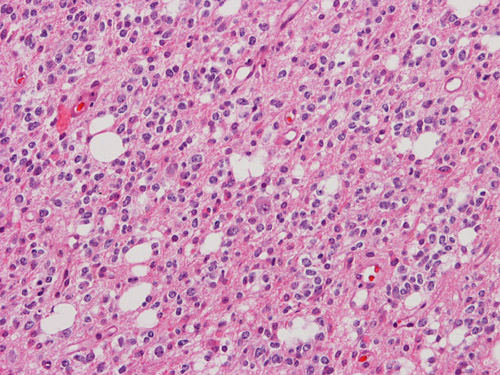
6. Which of the following features will be helpful in the diagnosis and prognostic assessment of the tumor illustrated below which is obtained from the frontal lobe of a 32 year-old man? Answer
Please click on icon to see full sized image.
A. Positive immunoreactivity for glial fibrillary acidic protein (GFAP)
B. Mutation of isocitrate dehydrogenase (IDH) 1 and lost of chromosome 1p and 19q
C. Weakly positive for synaptophysin staining
D. Demonstration of BRAF-KIAA1549 fusion
E. Demonstration of gemistocytic astrocytes
Kar-Ming Fung, M.D., Ph.D.
Last updated: October, 20. 2011.
Dept. of Pathology, University of Oklahoma Health Sciences Center, Oklahoma City, OK.
1. What is the histologic grade of diffuse astrocytoma in the WHO classification system? Answer
A. I.
B. II.
C. III.
D. IV.
E. V.
2. Which of the following morphologic features is sufficient to make a diagnosis of glioblastoma when the other features are compatible with such a diagnosis? Answer
A. Increased mitosis.
B. Endothelial proliferation.
C. Patchy distribution of positive immunoreactivity for glial fibrillary acidic protein (GFAP).
D. Increased nulcear pleomorphism.
E. Presence of sarcomatous component that is non-immunoreactive to glial fibrillary acidic protein (GFAP).
3. Which of the following features is true about glioblastomas? Answer
A. Secondary glioblastomas develop in older patients compared to primary glioblastoma.
B. T53 mutation is uncommon.
C. Giant cell glioblastoma carries a worse prognosis.
D. The 5 years survival rate for glioblastoma patient of all ages is about 20%..
E. Epidermal growth factor receptor (EGFR) amplication is common in older patients.
Kar-Ming Fung, M.D., Ph.D.
Last updated: November, 20, 2011.
Dept. of Pathology, University of Oklahoma Health Sciences Center, Oklahoma City, OK.
1. A 65 year-old man was admitted to the emergency room because of left side weakness. On neurologic examination, there was also paralysis of the left face. Ocular movement of both eyes, however, were normal. These symptoms were most likely resulted from blockage of which of the following arteries? Answer
A. Anterior communicating artery
B. Left anterior cerebral artery
C. Right anterior cerebral artery
D. Left middle cerebral artery
E. Right middle cerebral artery
2. A 45 year-old man with history of poorly controlled hypertension was admitted to the emergency by his friends. According to his friends, the patient has just collapsed in a banquet. On neurologic examination, the patient has unclear mental status but he was able to follow simple commands. There was significant weakness of his left upper and lower extremities. What is the most likely finding on CT scan? Answer
A. Hemorrhage in his right side basal ganglia with extension to the internal capsule
B. Hemorrhage in his right side basal ganglia with extension to the internal capsule
C. Hemorrhage in his hypothalamus with extension to the anterior commissure
D. Subdural hematoma in the frontal tip
E. Hemorrhage in the cerebellum
3. Bilateral infarction of the primary visual cortex will lead to which of the following syndrome? Answer
A. Sjögren syndrome
B. Anton syndrome
C. Parinaud syndrome
D. Susac syndrome
E. Wallenberg syndrome
4. Which of the following is not true about venous infarction due to thrombosis in the vein? Answer
A. Thrombosis of the of the cavernous sinus, sphenoid sinus, and lateral sinus is a common mechanism
B. It affects only elderly patients
C. It is common in cerebral amyloid angiopathy
D. Thrombolytic therapy will not lead to expansion of the hemorrhage
E. Pregnancy and use of oral conctraceptives are known risk factors
5. Foamy macrophages cannot be highlighted by which of the following techniques? Answer
A. Immunohistochemistry for CD68
B. Immunohistochemistry for CD163
C. Periodic acid Schiff (PAS) stain
D. Luxol fast blue-cresyl violet
6. This image is taken from the occipital lobe. What is the structure in between the two arrows? Answer
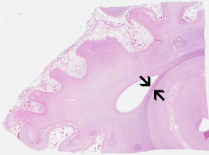
Click on icon to see enlarged image, Link here for online slide
A. Fimbria
B. Anterior commissure
C. Posterior commissure
D. Indusium griseum
E. Tapetum
Kar-Ming Fung, M.D., Ph.D.
Last updated: December, 20, 2011.
Dept. of Pathology, University of Oklahoma Health Sciences Center, Oklahoma City, OK.
1. Which of the followings is not true about meningioma? Answer
A. High mitotic rate (4 per 10 high power fields) is one of the diagnostic criteria for atypical meningioma (WHO grade II).
B. Brain invasion is one of the diagnostic criteria for atypical meningioma (WHO grade II).
C. Invasion into adjacent bone is one of the diagnostic criteria for atypical meningioma (WHO grade II).
D. Papillary meningioma and rhabdoid meningioma are anaplastic meningiomas (WHO grade III).
E. Meningiomas are positive for epithelial membrane antigen (EMA).
2. Which of the followings is not true about meningioma? Answer
A. Meningiomas are negative for cytokeratin.
B. S100 protein may have a patchy immunoreactivity in meningiomas.
C. Psammomatous meningiomas are WHO grade I tumor if there is no histologic features indicative of aggressive behavior or malignancy behavior.
D. Interdigitating cytoplasmic process is one of the ultrastructural features of meningiomas.
E. Meningiomas are typically contrast enhancing.
3. Which of the followings is not true about secretory meningioma? Answer
A. These tumors contain gland-like structures with eosinophilic, periodic acid Schiff (PAS) stain positive material.
B. Keratin and carcinoembryonic antigen (CEA) are typically positive around these vacuoles.
C. Histologically the gland-like structures may suggest metastatic carcinoma.
D. They rarely exist in pure form and classic meningotheliomatous areas are typically present.
E. Secretory meningiomas are seldom associated with impressive edema.
4. Which of the followings is not true about chordoid meningioma and chordoma? Answer
A. Both of them typically have a bluish, myxomatous to chondroid background that is strongly positive for Alcian blue.
B. Both of them can be positive for cytokeratin and epithelial membrane antigen (EMA).
C. Both of them can be positive for S100 protein and immunoreactivity is typically patchy.
D. Both of them are typically dural based.
E. Chordoma but not chordoid meningioma is positive for Brachyury.
5. Which of the following is true about meningioma in children? Answer
A. Meningiioma is a common tumor in children.
B. These tumors occur more often in an infratentorial, intraventricular, or intraparenchymal location than those in adults.
C. These tumors tend to be more aggressive and recur more frequently.
D. They are more often associated with neurofibromatosis 2 (NF2).
E. Meningioma and meningioangiomatosis are different entities and different in biological behavior.
Kar-Ming Fung, M.D., Ph.D.
Last updated: December, 20, 2014.
Dept. of Pathology, University of Oklahoma Health Sciences Center, Oklahoma City, OK.
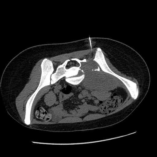
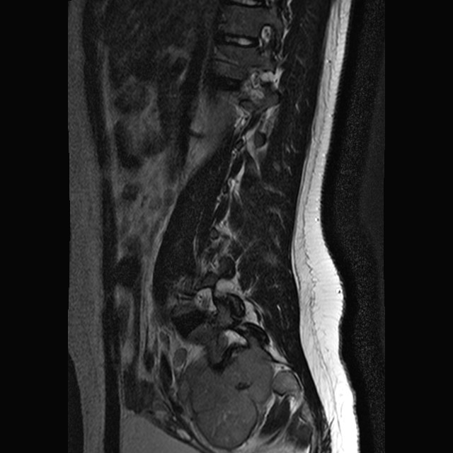
1. The patient was a 3 year-old girl with the chief complain of left lower extremity that extended to the right side. Imaging demonstrated a large sacral mass that mainly affects her left side. These features are not sufficient for a definitive diagnosis but which of the following be the most likely diagnosis? Answer
| CT taken at the time
of the needle biopsy (needle is shown here) |
MRI | FNA Diff Quick Stain |
Permanent
section Hematoxylin & Eosin Stain |
 |
 |
 |
 |
A. Osteosarcoma
B. Ependymoma, WHO grade II
C. Atypical teratoid/rhabdoid tumor (AT/RT)
D. Ewing's sarcoma
E. Metastatic Merkel cell carcinoma
2. Deletion or mutation of hSNF5/SMARCB1/INI1 has not been described in which of the following tumors? Answer
A. Medulloblastoma
B. Renal medullary carcinoma
C. Epithelioid sarcoma
D. Cribiform neuroepithelial tumor of the ventricle
E. Malignant rhabdoid tumor
Kar-Ming Fung, M.D., Ph.D.
1. Which of the following is the most common location of elastofibroma? Answer
A. Toes and fingers
B. Deep soft tissue on extremities
C. Around large joints such as the knee and hip joints
D. Subcutaneous location particularly on the back
E. Lower portion of scapula and chest wall
2. Elastofibroma are more common in man? Answer
A. True
B. False
3. Elastofibroma is seen most commonly in which of the following age group? Answer
A. Neonates and infants
B. Children and adolescents
C. Young adults
D. Elderly adults
Last updated: April, 7, 2017. Contributed by Kar-Ming Fung, M.D., Ph.D.
Dept. of Pathology, University of Oklahoma Health Sciences Center, Oklahoma City, OK, U.S.A.
Kar-Ming Fung, M.D., Ph.D.
Last updated: October, 20, 2011.
Dept. of Pathology, University of Oklahoma Health Sciences Center, Oklahoma City, OK.
1. Which of the following tumors are less likely to metastasize to bone? Answer
A. Leiomyosarcoma
B. Renal cell carcinoma
C. Adenocarcinoma arising in the lung
D. Invasive ductal carcinoma arising in the breast
E. Prostate carcinoma
2. Metastatic carcinoma, particularly at the early stage, is more common to be found in the axial skeleton? Answer
A. True
B. False
3. Which of the brain tumors has the highest tendency to metastasize to bone? Answer
A. Pleomorphic xanthoastrocytoma
B. Medulloblastoma
C. Glioblastoma
D. Pilocytic astrocytoma
E. Ependymoma arising in the brain
4. Which of the following combinations of immunohistochemistry would indicate a metastatic colorectal carcinoma? Answer
A. Cytokeratin 7 (-), Cytokeratin 20 (+), thyroid transcription factor 1 (-), villin (+)
B. Cytokeratin 7 (+), Cytokeratin 20 (-), thyroid transcription factor 1 (+), villin (-)
C. Cytokeratin 7 (+), Cytokeratin 20 (+), thyroid transcription factor 1 (-), villin (+)
D. Cytokeratin 7 (+), Cytokeratin 20 (-), synaptophysin (+)
E. Cytokeratin 7 (-), Cytokeratin 20 (-), Cytokerain 5/6 (+)
5. The overall rate of pathologic fracture associated with skeletal metastasis of prostate carcinoma is: Answer
A. Higher than other metastatic tumors.
B. The same as other metastatic tumors.
C. Lower than other metastatic tumor.
Kar-Ming Fung, M.D., Ph.D.
Last updated: December 3, 2011.
Dept. of Pathology, University of Oklahoma Health Sciences Center, Oklahoma City, OK.
1. Which of the following type of tumor is usually not osteolytic and less likely to be associtated with pathologic fracture when they metastasize to bone? Answer
A. Prostatic adenocarcinoma.
B. Clear cell renal cell carcinoma.
C. Papillary carcinoma of the thyroid.
D. Malignant melanoma.
E. Plasmacytoma/multiple myeloma.
2. Metastatic carcinoma does not always need extensive bone destruction to cause hypercalcemia? Answer
A. True.
B. False.
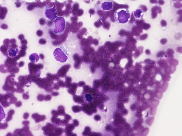
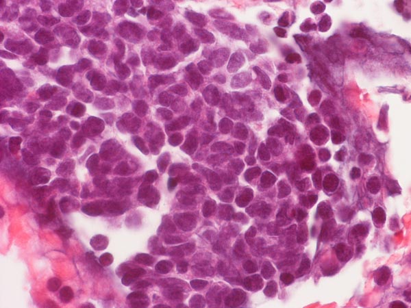
3. The patient is a 65 year-old man who complained of back pain. On his work up, a 5 cm osteolytic lesion is noted at the sacrum with a midline location and the tumor is roughly symmetrical. The patient has a history of renal cell carcinoma that was removed about 10 years ago. Histoloigic examination of the resected tumor yielded the following images. Which of the following sets of immunohistochemistry and/or molecular testing would allow you to correctly diagnose this tumor? Answer
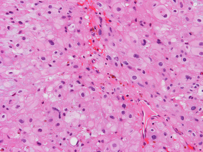
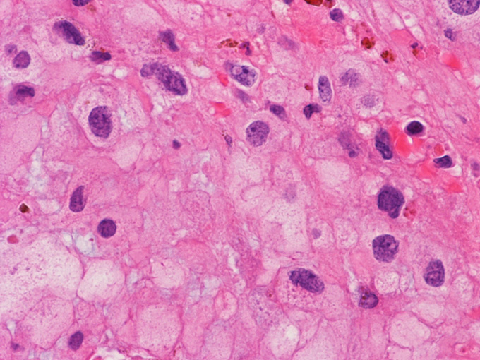
A. CD1a, CD163, S100, cytokeratin AE1/AE3.
B. Isocitrate dehydrogenase 1 and 2 gene (IDH2, IDH2), BRAF mutation and fusion, isochromosome 17.
C. Pax8, CD10, cytokeratin AE1/AE3.
D. Synaptophysin, S100, neurofilament.
E. S100, brachyury, CD10, cytokeratin AE1/AE3.
Kar-Ming Fung, M.D., Ph.D.
Last updated: November, 20, 2014.
Dept. of Pathology, University of Oklahoma Health Sciences Center, Oklahoma City, OK.
1. Which of the following statements is true? Answer
A. About 20% of all pituitary adenoma occurs in children.
B. The majority of tumors involving the sella in pediatric patients are craniopharyngioma.
C. Papillary craniopharyngioma is equally common in adult, adolescence, and children.
D. Endocrine dysfunction is uncommon in craniopharyngioma occuring in children.
E. Pituitary gigantism is not part of McCune Albright syndrome.
2. All of the following statements are true regarding the pharyngeal pituitary except which one? Answer
A. Its volume is about 1/1000 the volume of normal anterior pituitary.
B. The histologic component is identical to that of the anterior pituitary.
C. Pituitary adenoma can arise from pharyngeal pituitary.
D. It is located at the vellecula just anterior to the epiglottis.
3. Extracranial pituitary adenoma is most frequently found in which of the following locations? Answer
A. Sphenpid sinus.
B. Clivus.
C. Pharynx and/or nasopharynx.
D. Cavernous sinus.
4. Agenesis of anterior pituitary can occur in Pallister-Hall syndrome? Answer
A. True
B. False